. 2006 Aug;80(16):7939-51.
doi: 10.1128/JVI.00355-06.

Mutation of dileucine-like motifs in the human immunodeficiency virus type 1 capsid disrupts virus assembly, gag-gag interactions, gag-membrane binding, and virion maturation

Affiliations

Mutation of dileucine-like motifs in the human immunodeficiency virus type 1 capsid disrupts virus assembly, gag-gag interactions, gag-membrane binding, and virion maturation

Anjali Joshi et al. J Virol. 2006 Aug.

Abstract

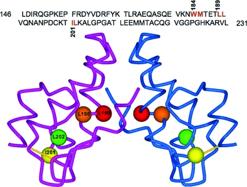
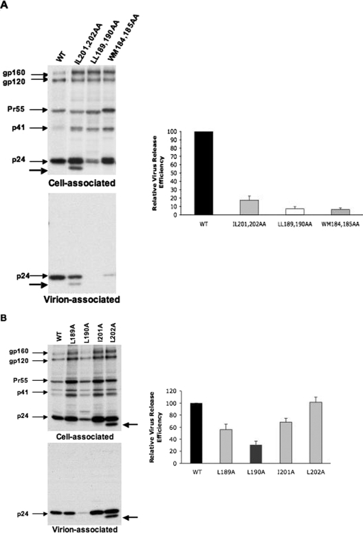
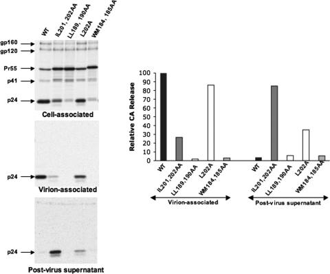
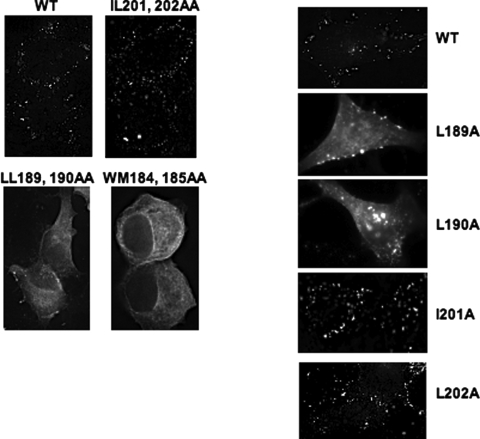
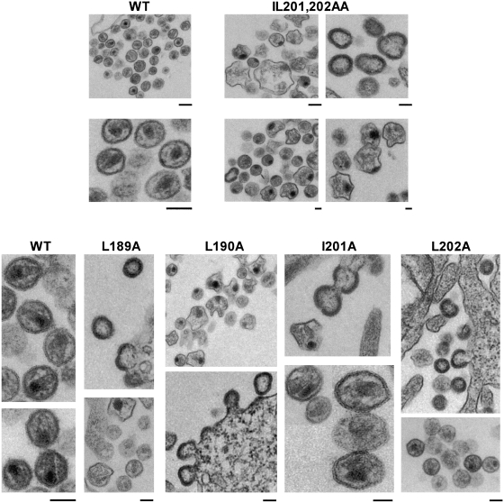
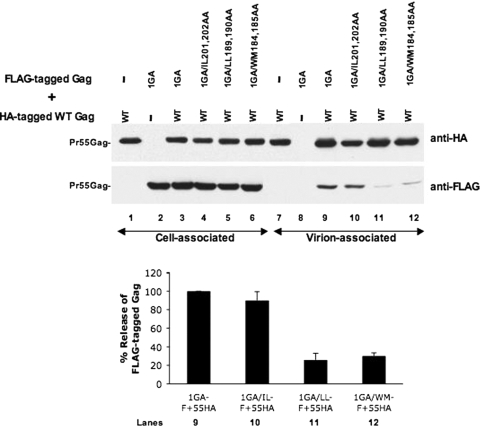
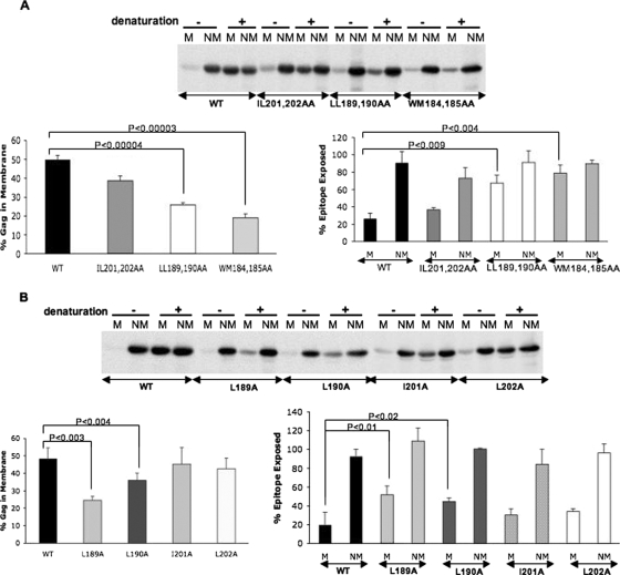
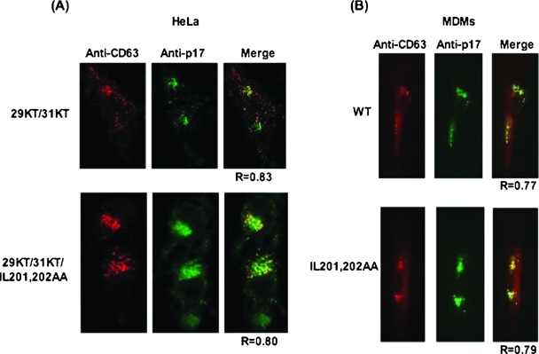
The human immunodeficiency virus type 1 (HIV-1) Gag precursor protein Pr55(Gag) drives the assembly and release of virus-like particles in the infected cell. The capsid (CA) domain of Gag plays an important role in these processes by promoting Gag-Gag interactions during assembly. The C-terminal domain (CTD) of CA contains two dileucine-like motifs (L189/L190 and I201/L202) implicated in regulating the localization of Gag to multivesicular bodies (MVBs). These dileucine-like motifs are located in the vicinity of the CTD dimer interface, a region of CA critical for Gag-Gag interactions during virus assembly and CA-CA interactions during core formation. To study the importance of the CA dileucine-like motifs in various aspects of HIV-1 replication, we introduced a series of mutations into these motifs in the context of a full-length, infectious HIV-1 molecular clone. CA mutants LL189,190AA and IL201,202AA were both severely impaired in virus particle production because of a variety of defects in the binding of Gag to membrane, Gag multimerization, and CA folding. In contrast to the model suggesting that the CA dileucine-like motifs regulate MVB targeting, the IL201,202AA mutation did not alter Gag localization to the MVB in either HeLa cells or macrophages. Revertants of single-amino-acid substitution mutants were obtained that no longer contained dileucine-like motifs but were nevertheless fully replication competent. The varied phenotypes of the mutants reported here provide novel insights into the interplay among Gag multimerization, membrane binding, virus assembly, CA dimerization, particle maturation, and virion infectivity.


Figures

FIG. 1.
FIG. 1.
Sequence and structure of the HIV-1 CA CTD. The amino acid sequence of the CA CTD is shown at the top. The CTD dimer interface residues W184/M185 and the putative di-Leu-like motifs L189/L190 and I201/L202 are indicated in red. At the bottom is shown a structural model of a dimer of the HIV-1 CA CTD (provided by S. R. Durell) with the locations of L189, L190, I201, and L202 indicated by the red, orange, yellow, and green balls, respectively. Residue L189 lies at the CA dimer interface; residues L190 and L202 are buried, while residue I201 is exposed. The structure was constructed with data from 1A43.pdb (51).
FIG. 2.
FIG. 2.
The CA mutants LL189,190AA and IL201,202AA are severely defective in virus assembly and release. HeLa cells were transiently transfected with WT pNL4-3 (WT) or derivatives containing the indicated (A) CA double or (B) CA single mutations. Transfected HeLa cells were metabolically labeled with [35S]Met/Cys, and virions were pelleted by ultracentrifugation. Cell and virion lysates were immunoprecipitated with HIV-Ig and analyzed by SDS-PAGE, followed by phosphorimager analysis. The positions of the Env precursor gp160, the mature surface Env glycoprotein gp120, the Gag precursor Pr55Gag, the Gag processing intermediate p41Gag, and p24 (CA) are shown. The ∼20-kDa protein product detected with the IL201,202AA and L202A mutants is indicated by an unmarked arrow. Virus release efficiency was calculated from at least three independent experiments as the amount of radiolabeled, virion-associated Gag as a fraction of the total (cell plus virion) radiolabeled Gag protein detected. Error bars show standard deviations.
FIG. 3.
FIG. 3.
Mutation of L202 leads to the release of free CA and CA-derived product. HeLa cells transfected with WT pNL4-3 or the indicated CA mutants were metabolically labeled with [35S]Met/Cys, and virions were pelleted by ultracentrifugation. Cell- and virion-associated material and the supernatant obtained from virus pelleting (postvirus supernatant) were immunoprecipitated with HIV-Ig and analyzed by SDS-PAGE, followed by phosphorimager quantification. Results from one representative experiment are shown; similar results were obtained in an independently repeated experiment.
FIG. 4.
FIG. 4.
Replication kinetics of CA mutants and isolation of viral revertants. (A) MT-4 T cells were transfected with either WT pNL4-3 or the indicated CA single mutants. Cells were split every 2 days; supernatant was reserved at each time point for RT analysis. Repassage of CA single mutants and isolation of viral revertants. (B) Jurkat and (C) MT-4 T-cell lines were infected with WT or CA mutant virus (normalized for RT activity) obtained from the MT-4 replication experiment shown in panel A. Culture supernatant was reserved every 2 days for RT analysis. Cell pellets were also harvested for DNA isolation, PCR amplification, and sequencing of the gag gene. P2 after the mutant name denotes passage 2. (D) Sequence profile of revertant viruses obtained from repassage of CA single mutants in Jurkat and MT-4 cells. The codons found in WT pNL4-3, the mutant derivatives, and the PCR-amplified viral DNA are indicated; the encoded amino acid is indicated in parentheses. For L202, no revertants were obtained (none).
FIG. 5.
FIG. 5.
Subcellular localization of CA-mutant Gag. HeLa cells were transfected with WT pNL4-3 or the indicated CA-mutant derivatives. Cells were fixed 24 h posttransfection and stained with anti-HIV-1 p17 (MA) Ab followed by fluorescence microscopy with a DeltaVision RT microscope.
FIG. 6.
FIG. 6.
EM analysis of CA-mutant virions. HeLa cells were transfected with WT pNL4-3 or the indicated CA-mutant derivatives. Cells were fixed at 2 days posttransfection and observed by transmission EM. The bar under each panel represents a 100-nm scale.
FIG. 7.
FIG. 7.
Analysis of the rescue of CA mutant Gag into VLPs upon coexpression with WT Gag. HeLa cells were transfected with FLAG-tagged versions of either 1GA/pNL4-3 or 1GA/pNL4-3 derivatives containing the indicated CA double mutations. Rescue of the nonmyristylated (1GA) Gag protein into VLPs was measured by cotransfection with WT HA-tagged Gag. Cell and virus lysates were subjected to SDS-PAGE, followed by immunoblotting with anti-HA or anti-FLAG Ab. Quantification of Pr55Gag bands was performed with the Alpha Innotech digital imaging system. Virus release efficiency was calculated as the percent release of FLAG-tagged 1GA Gag, with the amount of FLAG-tagged 1GA Gag containing WT CA rescued into VLPs by HA-tagged Gag being given a relative value of 100%. Lane numbers under the graph correspond to the respective lanes in the anti-FLAG Western blot. In the graph, 1GA-F, 1GA/IL-F, 1GA/LL-F, and 1GA/WM-F refer to FLAG-tagged 1GA Gag proteins containing WT, IL201,202AA, LL189,190AA, and WM184,185AA CA, respectively. 55HA denotes HA-tagged WT Gag. Data represent averages from at least three independent experiments ± the standard deviations.
FIG. 8.
FIG. 8.
Effects of CA mutations on membrane binding and Gag multimerization. HeLa cells were transfected with pNL4-3/PR versions of the (A) CA double or (B) CA single mutants. Transfected cells were pulse-chase labeled with [35S]Met/Cys and sonicated, and postnuclear supernatants were subjected to membrane flotation centrifugation on sucrose gradients (see Materials and Methods). The membrane (M) and nonmembrane (NM) fractions were isolated and immunoprecipitated with HIV-Ig either with (+) or without (−) prior denaturation. Pr55Gag bands were quantified by phosphorimager analysis. The graphs in the lower left of panels A and B indicate membrane binding efficiency. Percent epitope exposure (lower right), which is a measurement of higher-order Gag multimerization (39), was determined by calculating the percentage of Gag recovered by immunoprecipitation from nondenatured relative to denatured fractions. Note that epitope masking is observed predominantly with membrane-associated Gag. P values represent significant differences between the WT and mutant Gag proteins as determined by the Student t test.
FIG. 9.
FIG. 9.
MVB localization of the IL201,202AA CA mutant. (A) HeLa cells were transfected with pNL4-3/29KT/31KT (top) or with the derivative containing the IL201,202AA CA mutation (bottom). (B) WT NL4-3 or the IL201,202AA CA-mutant derivative was expressed in primary human MDMs (Materials and Methods). Cells were fixed 24 h posttransfection, permeabilized, and stained with anti-HIV-1 p17 (MA) (green) and anti-CD63 (red) Abs, followed by microscopy with a DeltaVision RT microscope. The softWoRx colocalization module was used to determine costaining between Gag and CD63 on an average of 8 to 10 cells. One representative field for each is depicted. When data were compiled and averaged for all of the cells analyzed, the Gag/CD63 colocalization in the context of WT CA was R = 0.82 ± 0.05 in HeLa cells and R = 0.7028 ± 0.17 in MDMs; for the IL201,202AA CA mutant, it was R = 0.80 ± 0.04 in HeLa cells and 0.6826 ± 0.102 in MDMs. R = Pearson coefficient of correlation.

Similar articles

Cited by

References

    1. Bonifacino, J. S., and L. M. Traub. 2003. Signals for sorting of transmembrane proteins to endosomes and lysosomes. Annu. Rev. Biochem. 72:395-447. - PubMed
    1. Burniston, M. T., A. Cimarelli, J. Colgan, S. P. Curtis, and J. Luban. 1999. Human immunodeficiency virus type 1 Gag polyprotein multimerization requires the nucleocapsid domain and RNA and is promoted by the capsid-dimer interface and the basic region of matrix protein. J. Virol. 73:8527-8540. - PMC - PubMed
    1. Campbell, S., and A. Rein. 1999. In vitro assembly properties of human immunodeficiency virus type 1 Gag protein lacking the p6 domain. J. Virol. 73:2270-2279. - PMC - PubMed
    1. Campbell, S., and V. M. Vogt. 1995. Self-assembly in vitro of purified CA-NC proteins from Rous sarcoma virus and human immunodeficiency virus type 1. J. Virol. 69:6487-6497. - PMC - PubMed
    1. Cimarelli, A., S. Sandin, S. Hoglund, and J. Luban. 2000. Basic residues in human immunodeficiency virus type 1 nucleocapsid promote virion assembly via interaction with RNA. J. Virol. 74:3046-3057. - PMC - PubMed

Publication types

MeSH terms

Substances

LinkOut - more resources