. 2006 Aug;80(16):7995-8005.
doi: 10.1128/JVI.00734-06.

PML contributes to a cellular mechanism of repression of herpes simplex virus type 1 infection that is inactivated by ICP0

Affiliations

PML contributes to a cellular mechanism of repression of herpes simplex virus type 1 infection that is inactivated by ICP0

Roger D Everett et al. J Virol. 2006 Aug.

Abstract

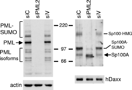
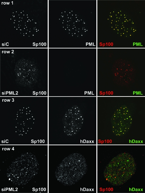
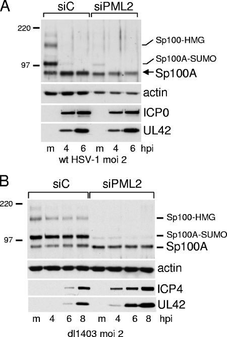
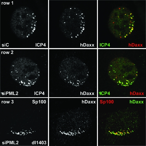
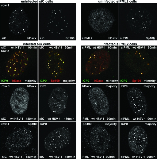
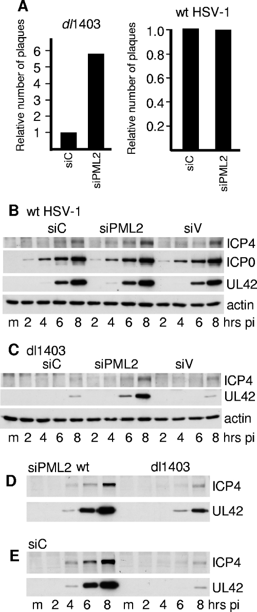
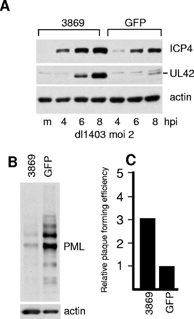
Promyelocytic leukemia (PML) nuclear bodies (also known as ND10) are nuclear substructures that contain several proteins, including PML itself, Sp100, and hDaxx. PML has been implicated in many cellular processes, and ND10 are frequently associated with the replicating genomes of DNA viruses. During herpes simplex virus type 1 (HSV-1) infection, the viral regulatory protein ICP0 localizes to ND10 and induces the degradation of PML, thereby disrupting ND10 and dispersing their constituent proteins. ICP0-null mutant viruses are defective in PML degradation and ND10 disruption, and concomitantly they initiate productive infection very inefficiently. Although these data are consistent with a repressive role for PML and/or ND10 during HSV-1 infection, evidence in support of this hypothesis has been inconclusive. By use of short interfering RNA technology, we demonstrate that depletion of PML increases both gene expression and plaque formation by an ICP0-negative HSV-1 mutant, while having no effect on wild-type HSV-1. We conclude that PML contributes to a cellular antiviral repression mechanism that is countered by the activity of ICP0.


Figures

FIG. 1.
FIG. 1.
Effect of shPML2 expression on PML protein levels and SUMO modification of Sp100 in retrovirally transduced HF cells. Whole-cell extracts from 5 × 10⁴ untreated cells were analyzed by Western blotting for PML, Sp100, hDaxx, and actin. The left panels show PML protein levels in siC, siPML2, and siV cells compared to actin. The major PML isoform is shown by an arrow, with the presumed SUMO-modified species migrating more slowly. The faster-migrating bands detected by 5E10 are definitively identified as lower-molecular-weight PML isoforms by this approach. The lower right panel shows that the levels of hDaxx are unaffected by ablation of PML. The upper right panel illustrates that there are several differences in the pattern of Sp100 isoforms in PML-depleted cells, as discussed in detail in the text. Numbers between the panels show molecular masses in kilodaltons.
FIG. 2.
FIG. 2.
Distributions of PML, Sp100, and hDaxx in siPML2 cells and control fibroblasts. Rows 1 and 2: siC (row 1) and siPML2 (row 2) cells were costained for PML (5E10; green) and Sp100 (SpGH; red). The images were captured using identical microscope settings. Rows 3 and 4: siC (row 3) and siPML2 (row 4) cells were costained for hDaxx (r1866; green) and Sp100 (rat26; red).
FIG. 3.
FIG. 3.
(A) ICP0 has no effect on unmodified Sp100 during wt HSV-1 infection of cells lacking PML. siC and siPML2 cells were infected with wt HSV-1 (MOI of 2), and then samples were harvested at 4 and 6 h after infection for analysis by Western blotting for Sp100, actin, ICP0, and UL42 as indicated. (B) The loss of the higher-molecular-weight isoforms of Sp100 during wt HSV-1 infection does not occur in ICP0-null mutant dl1403-infected cells. siC and siPML2 cells were infected with dl1403 at an MOI of 2, and samples were taken at the indicated times after infection for analysis by Western blotting for Sp100, actin, ICP4, and UL42. hpi, hours postinfection. Numbers at left are molecular masses in kilodaltons. m, mock infected.
FIG. 4.
FIG. 4.
Redistribution of ND10 proteins in PML-depleted and control cells at the edges of plaques of ICP0-null mutant virus dl1403. siC cells (rows 1 and 3) and siPML2 cells (rows 2 and 4) were infected with dl1403 at an MOI of 0.1 and then stained the following day for ICP4 (58S; green) and PML (r8; red) (rows 1 and 2) or ICP4 (58S; green) and Sp100 (SpGH; red). Cells in the early stages of infection at the edges of plaques displaying the characteristic asymmetric distributions of early ICP4 foci were selected for imaging without prior visual inspection of the PML or Sp100 signals. The image capture settings for the control cells (rows 1 and 3) were reused without adjustment for the siPML2 cells (rows 2 and 4).
FIG. 5.
FIG. 5.
Redistribution of ND10 proteins in PML-depleted and control cells at the edges of plaques of ICP0-null mutant virus dl1403. siC (row 1) and siPML2 (row 2) cells were infected with dl1403 at an MOI of 0.1 and then stained the following day for ICP4 (58S; green) and hDaxx (r1866; red). Cells in the early stages of infection at the edges of plaques displaying the characteristic asymmetric distributions of early ICP4 foci were selected for imaging without prior visual inspection of the hDaxx signal. The image capture settings for the control cells were reused without adjustment for the siPML2 cells. Row 3 shows a similarly infected siPML2 cell costained for Sp100 (rat26; red) and hDaxx (r1866; green). Although not stained for a viral antigen, viral plaques in this sample could be readily identified by the characteristic changes that occur in the distributions of Sp100 and hDaxx compared to neighboring uninfected cells (compare with Fig. 2, row 4).
FIG. 6.
FIG. 6.
ICP0 initially colocalizes with and then disperses Sp100 and hDaxx in both siC and siPML2 cells. siC and siPML2 cells were infected with wt HSV-1 (MOI of 2), and samples were analyzed by immunofluorescence after 90 and 180 minutes of infection. The panels are appropriately labeled to indicate the cell type, the time point, and the staining regimen used. Row 2 shows individual cells dual labeled for the indicated proteins. Rows 1, 3, and 4 show the separated red and green channels for either single siC cells (left of panels) or siPML2 cells (right of panels). Row 1 shows control uninfected siC and siPML2 cells, and rows 2 to 4 show infected siC and siPML2 cells as indicated.
FIG. 7.
FIG. 7.
(A) Relative efficiencies of wt and ICP0-null mutant HSV-1 dl1403 plaque formation in cells transduced with retroviruses expressing anti-PML or control shRNAs. The increase of dl1403 titers in siPML2 compared to siC cells varied from 2.4- to 10.0-fold, the average of eight experiments being 5.8-fold. In a typical example of an experiment, the titers of a stock of dl1403 in siC, siPML2, and U2OS cells were 9.2 × 10⁴, 5.2 × 10⁵, and 4.8 × 10⁷, respectively. The titers were determined by a series of threefold dilutions from an appropriate starting point. For wt HSV-1, the average relative titers on siPML2 cells compared to siC cells over three experiments were 0.82, 1.2, and 0.92 (average, 0.98). In a typical example of an experiment, the titers of a stock of wt HSV-1 in siC, siPML2, and U2OS cells were 6.7 × 10⁸, 4.9 × 10⁸, and 5.8 × 10⁸, respectively. (B to E). Increase in gene expression of ICP0-null but not wt HSV-1 in cells depleted for PML. siC, siPML2, and siV cells were seeded into 24-well dishes. The following day, the cells in one well of each cell type were resuspended in trypsin and counted. Cells were then infected at an MOI of 2 with either wt HSV-1 or dl1403, and then samples were harvested at 2, 4, 6, and 8 h after infection using volumes of gel loading buffer to give equal total cell protein concentrations. Actin levels and expression of ICP4 and UL42 (and ICP0 in the wt HSV-1 samples) were analyzed by Western blotting. (B) wt HSV-1 infection of siC, siPML2, and siV cells. (C) dl1403 infection of siC, siPML2, and siV cells. (D) The wt HSV-1 and dl1403 samples from the siPML2 infection analyzed on the same gel. (E) The wt HSV-1 and dl1403 samples from the siC infection analyzed on the same gel. pi, postinfection. m, mock infected.
FIG. 8.
FIG. 8.
Enhancement of dl1403 gene expression and plaque formation in cells expressing a different anti-PML shRNA. HF cells were transduced with lentiviruses expressing control GFP or anti-PML shRNA 3869. m, mock infected. (A) Cells were infected with dl1403 at an MOI of 2, and samples were harvested at the indicated time points. Expression of ICP4, UL42, and actin was detected by Western blotting. (B) PML expression in HF-3869 and HF-GFP cells compared to actin. (C) Relative plaque-forming efficiency of dl1403 in HF-3869 cells, relative to that in HF-GFP cells. The increase in plaque formation in HF-3869 cells averaged 3.0-fold from four replicate experiments, with a range of 2.0 to 3.6.

Similar articles

Cited by

References

    1. Ahn, J. H., and G. S. Hayward. 2000. Disruption of PML-associated nuclear bodies by IE1 correlates with efficient early stages of viral gene expression and DNA replication in human cytomegalovirus infection. Virology 274:39-55. - PubMed
    1. Bernardi, R., and P. P. Pandolfi. 2003. Role of PML and the PML-nuclear body in the control of programmed cell death. Oncogene 22:9048-9057. - PubMed
    1. Borden, K. L. 2002. Pondering the promyelocytic leukemia protein (PML) puzzle: possible functions for PML nuclear bodies. Mol. Cell. Biol. 22:5259-5269. - PMC - PubMed
    1. Canning, M., C. Boutell, J. Parkinson, and R. D. Everett. 2004. A RING finger ubiquitin ligase is protected from autocatalyzed ubiquitination and degradation by binding to ubiquitin-specific protease USP7. J. Biol. Chem. 279:38160-38168. - PubMed
    1. Chee, A. V., P. Lopez, P. P. Pandolfi, and B. Roizman. 2003. Promyelocytic leukemia protein mediates interferon-based anti-herpes simplex virus 1 effects. J. Virol. 77:7101-7105. - PMC - PubMed

Publication types

MeSH terms

LinkOut - more resources