. 2006 Aug;80(16):8006-18.
doi: 10.1128/JVI.00743-06.

Evidence for a role of the cellular ND10 protein PML in mediating intrinsic immunity against human cytomegalovirus infections

Affiliations

Evidence for a role of the cellular ND10 protein PML in mediating intrinsic immunity against human cytomegalovirus infections

Nina Tavalai et al. J Virol. 2006 Aug.

Abstract

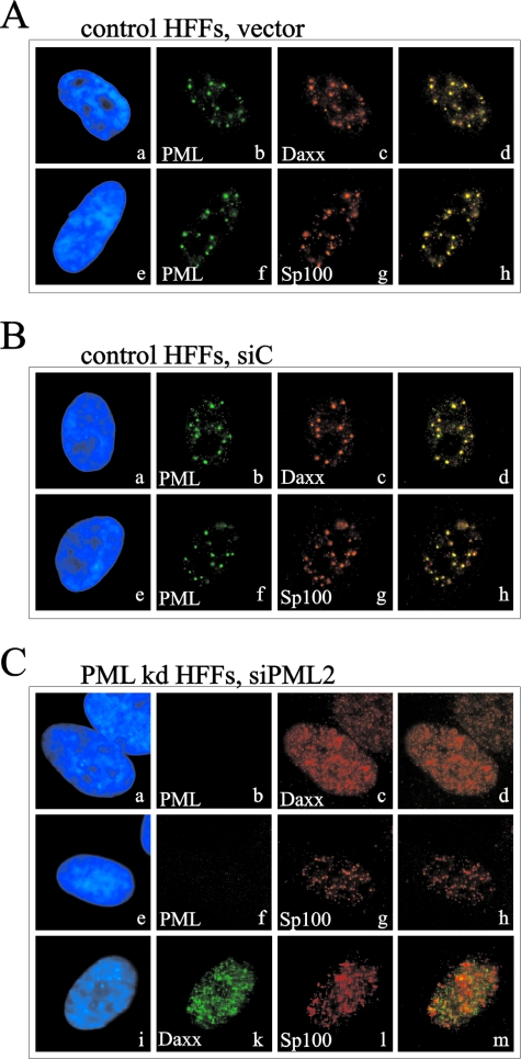
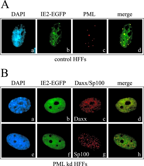
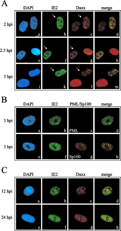
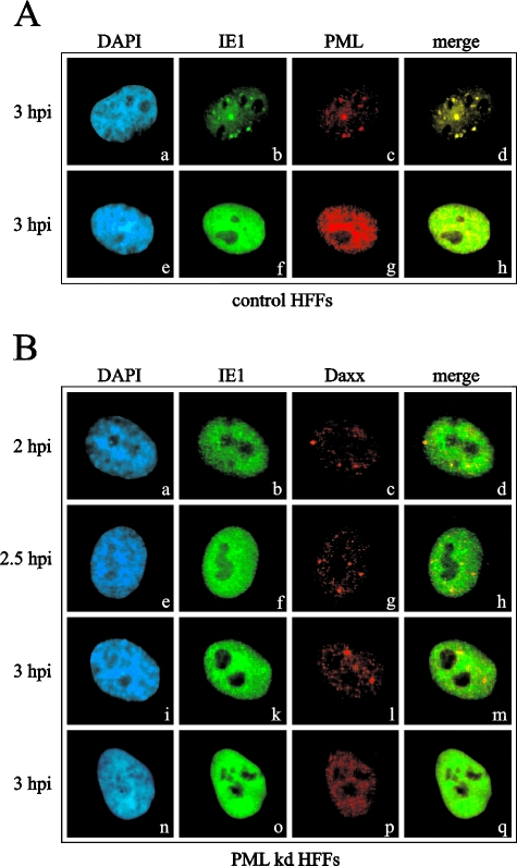
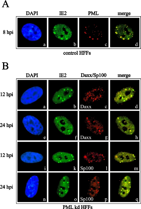
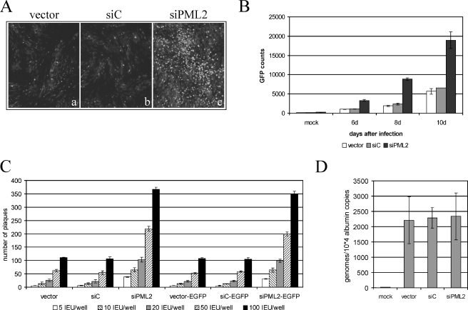
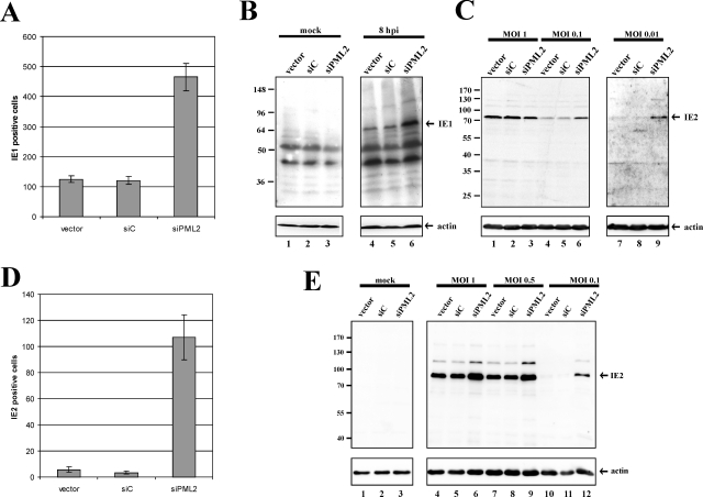
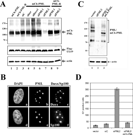
Several viruses, including human cytomegalovirus (HCMV), encode proteins that colocalize with a cellular subnuclear structure known as ND10. Since only viral DNA deposited at ND10 initiates transcription, ND10 structures were hypothesized to be essential for viral replication. On the other hand, interferon treatment induces an up-regulation of ND10 structures and viruses have evolved polypeptides that disperse the dot-like accumulation of ND10 proteins, suggesting that ND10 could also be part of an intrinsic defense mechanism. In order to obtain evidence for either a proviral or an antiviral function of ND10, we generated primary human fibroblasts with a stable, short interfering RNA-mediated knockdown (kd) of PML. In these cells, other ND10-associated proteins like hDaxx showed a diffuse nuclear distribution. Interestingly, we observed that HCMV infection induced the de novo formation of ND10-like hDaxx and Sp100 accumulations that colocalized with IE2 and were disrupted, in the apparent absence of PML, in an IE1-dependent manner during the first hours after infection. Furthermore, infection of PML-kd cells with wild-type HCMV at a low multiplicity of infection resulted in enhanced replication. In particular, a significantly increased plaque formation was detected, suggesting that more cells are able to support initiation of replication in the absence of PML. While there was no difference in viral DNA uptake between PML-kd and control cells, we observed a considerable increase in the number of immediate-early (IE) protein-positive cells, indicating that the depletion of PML augments the initiation of viral IE gene expression. These results strongly suggest that PML functions as part of an intrinsic immune mechanism against cytomegalovirus infections.


Figures

FIG. 1.
FIG. 1.
Localization of target sequences for siRNAs against PML relative to the localization of exons and protein domains (data taken from reference 34). All PML isoforms share a common N terminus but differ in their C termini due to alternative usage of 3′ exons. Protein domains of PML are given in the upper part of the figure. R, RING finger; B1 and B2, B boxes; CC, coiled-coil domain; SUMO, SUMOylation sites. The lower part illustrates the exon assembly of various PML isoforms. Positions within PML that are targeted by the siRNAs siPML1 and siPML2 are indicated by arrows.
FIG. 2.
FIG. 2.
Detection of endogenous PML by Western blot analysis of cell lysates derived from primary human fibroblasts transduced with various siRNA expression vectors as indicated. (A) Upper panel: monoclonal anti-PML antibody 5E10 was used to detect PML; lower panel: beta-actin was detected as a loading control. (B) Comparison of the reactivities of anti-PML monoclonal antibody 5E10 and anti-PML rabbit serum H-238 using cell lysates from primary human fibroblasts transduced with pSIREN-RetroQ vectors as indicated. Lanes 1 to 3, upper panel: monoclonal 5E10; lower panel: beta-actin antibody. Lanes 4 to 6, upper panel: anti-PML rabbit serum H-238; lower panel: anti-Daxx rabbit serum M-117. The localization of PML isoforms is indicated on the right of each panel; the asterisks indicate a nonspecific reaction of anti-PML antibody 5E10. Numbers at left of each panel are molecular masses in kilodaltons.
FIG. 3.
FIG. 3.
Stable knockdown of PML in primary human fibroblasts leads to a dispersal of ND10 domains with a dissociation of hDaxx and Sp100. The figure shows detection of ND10 structures in primary human fibroblasts stably transduced with various siRNA vectors by indirect immunofluorescence analysis. The following antibodies were used for staining: anti-PML monoclonal antibody PG-M3 (b and f), anti-hDaxx polyclonal serum M-117 (c), and anti-Sp100 polyclonal serum H-60 (g); the costaining in panel C, subpanels i to m, was performed using anti-hDaxx monoclonal antibody MCA2143 (k) together with anti-Sp100 serum H-60 (l). DAPI (4′,6′-diamidino-2-phenylindole) staining of the respective cell nuclei is shown in subpanels a, e, and i; subpanels d, h, and m show merged images of PML/hDaxx, PML/Sp100, or hDaxx/Sp100 staining, respectively. (A) pSIREN-RetroQ-transduced control HFFs. (B) pSIREN-RetroQ-siC-transduced control fibroblasts. (C) pSIREN-RetroQ-siPML2-transduced PML-kd fibroblasts.
FIG. 4.
FIG. 4.
Localization of IE2-p86 after transient expression in PML knockdown or control fibroblasts. Control HFFs (A) or siPML2-expressing PML-kd fibroblasts (B), grown on coverslips, were transfected with a eukaryotic expression vector encoding IE2-p86 fused to EGFP. Thereafter, indirect immunofluorescence analyses were carried out. IE2-p86 was detected via EGFP autofluorescence (A, subpanel b; B, subpanels b and f); endogenous ND10 proteins were visualized via anti-PML monoclonal antibody PG-M3 (A, subpanel c) or via the polyclonal rabbit sera M-117 against hDaxx (B, subpanel c) and H-60 against Sp100 (B, subpanel g). DAPI, 4′,6′-diamidino-2-phenylindole.
FIG. 5.
FIG. 5.
Analysis of the intranuclear localization of IE2 and cellular ND10 proteins in PML knockdown cells after infection with wild-type HCMV (AD169). (A and B) Subnuclear localization of IE2 together with hDaxx (A) or PML/Sp100 (B) during the first 2 to 3 hpi of PML-kd cells. Arrows in panel A indicate HCMV-infected cells. IE2 was detected by monoclonal antibody SMX; hDaxx, PML, or Sp100 was detected by polyclonal antiserum M-117, H-238, or H-60, respectively. IE2 is detectable in punctate foci which colocalize with reorganized ND10-like structures containing hDaxx (A; compare infected with noninfected cells) and Sp100 (B). (C) Subnuclear localization of IE2 and hDaxx at 12 or 24 hpi. IE2 was detected by monoclonal antibody SMX; hDaxx was stained using polyclonal serum M-117.
FIG. 6.
FIG. 6.
Analysis of the subcellular localization of IE1 and the ND10 components PML and hDaxx during the time course of infection in PML-kd and control fibroblasts. Control fibroblasts, transduced with pSIREN-RetroQ (A), and siPML2-transduced cells (B) grown on coverslips were infected with AD169 at an MOI of 1 and harvested during the first hours postinfection as indicated. Indirect immunofluorescence analysis was performed to investigate the intranuclear localization of IE1, hDaxx, and PML by utilization of the anti-IE1 antibody p63-27 as well as the polyclonal antisera H-238 and M-117 for detection of PML and hDaxx, respectively. DAPI, 4′,6′-diamidino-2-phenylindole.
FIG. 7.
FIG. 7.
Subnuclear distribution of PML, hDaxx, and Sp100 in PML-deficient and control cells infected with the IE1 deletion virus CR208. Control HFFs, transduced with pSIREN-RetroQ (A), and siPML2-transduced PML-kd cells (B) grown on coverslips were infected with the IE1 mutant virus CR208 at an MOI of 1. In subsequent immunofluorescence experiments the formation of distinct PML, hDaxx, and Sp100 foci was examined at the indicated time points after infection using the anti-PML antibody PG-M3, the anti-Daxx antibody MCA2143, and the anti-Sp100 antiserum H-60. Infected cells were identified by staining of the IE2 protein with the monoclonal antibody SMX (B, subpanels k and o) or the polyclonal antiserum pHM178 (A, subpanel b; B, subpanels b and f). DAPI, 4′,6′-diamidino-2-phenylindole.
FIG. 8.
FIG. 8.
Quantification of HCMV replication in retrovirally transduced HFFs expressing siRNAs. (A and B) Retrovirally transduced HFFs (vector, siC, and siPML2) were infected with recombinant HCMV AD169-GFP expressing green fluorescent protein. Panel A shows the GFP fluorescence 10 days after infection of the indicated HFFs (MOI of 0.02). Panel B shows a quantification of the GFP fluorescence by automated fluorometry at days 6, 8, and 10 after infection with HCMV AD169-GFP. (C) Quantification of the number of plaques by standard plaque assays 7 days after infection of transduced HFFs (vector, siC, siPML2, vector-EGFP, siC-EGFP, and siPML2-EGFP) with the indicated amount of HCMV strain AD169 (viral inoculum ranging from 5 to 100 IEU of virus per well of a six-well dish). (D) Quantitative real-time PCR for evaluation of the viral DNA load after infection of PML-kd cells in comparison to control fibroblasts. HFF cells as indicated were infected with AD169 at an MOI of 0.02. Then, DNA was extracted 24 hpi and the number of HCMV genome copies was determined by real-time PCR. The amplification of the cellular albumin gene was utilized as a standard for calculating the cell numbers.
FIG. 9.
FIG. 9.
Analysis of IE gene expression after infection of PML-kd and control cells with wild-type HCMV or the IE1 deletion mutant CR208. (A and D) PML-negative (siPML2) and control (vector, siC) HFFs, grown on coverslips in six-well dishes, were infected with 100 IEU/well of either HCMV strain AD169 (A) or IE1 deletion mutant CR208 (D), respectively. Cells were fixed at 24 hpi, and the number of IE-expressing cells was determined by indirect immunofluorescence analysis using either antibody p63-27 against IE1 (A) or antiserum anti-pHM178 against IE2 (D). (B, C, and E) Western blot analyses of IE gene expression in retrovirally transduced HFFs expressing the respective siRNAs infected with either HCMV AD169 (B and C) or IE1 deletion mutant CR208 (E). (B) HFFs were infected with HCMV AD169 at an MOI of 0.01. Lysates were harvested 8 hpi and analyzed by Western blotting for IE1 gene expression using monoclonal antibody p63-27. (C) HFFs were infected with HCMV AD169 at various MOIs as indicated, and cell extracts were harvested 24 hpi for the detection of the IE protein IE2 (anti-pHM178). (E) HFFs were infected with CR208 at various MOIs as indicated, and cell extracts were harvested 24 hpi for detection of the IE protein IE2 (anti-pHM178). Numbers at left of panels B, C, and E are molecular masses in kilodaltons.
FIG. 10.
FIG. 10.
Reconstitution of PML expression in siPML2 cells. (A) Verification of a PML construct which is resistant against degradation mediated by the PML-siRNA siPML2. HEK293T cells were cotransfected with the respective PML-expressing constructs (mCh-PML comprises the wild-type PML sequence; mCh-PML-R illustrates the degradation-resistant isoform) and a plasmid coding for FLAG-tagged UL69 as well as vectors containing the different siRNA sequences as indicated. Thereafter, cell extracts were analyzed for PML expression using the polyclonal rabbit serum H-238 (upper panel). FLAG-tagged pUL69 was detected by monoclonal antibody anti-FLAG M2 (middle panel). The detection of actin (lower panel) was used as a loading control. (B) Indirect immunofluorescence analysis for detection of PML in siPML2 cells after retroviral gene transfer of the mCh-PML fusion sequence. PML was visualized through autofluorescence of its mCherry moiety. hDaxx and Sp100 were stained using the polyclonal antisera M-117 and H-60, respectively. DAPI, 4′,6′-diamidino-2-phenylindole. (C) Detection of mCh-PML by Western blotting after transduction of siPML2 cells with the mCh-PML expression vector resulting in siPML2/mCh-PML cells. Endogenous PML (lane 1, vector) and reintroduced mCh-PML (lane 3, siPML2/mCh-PML) were detected with the help of the polyclonal antiserum H-238. siPML2 cells, used for transduction with mCh-PML, were analyzed in parallel (lane 2). (D) Analysis of IE gene expression after reintroduction of PML in PML kd cells. The different cell populations as indicated were infected with 25 IEU of HCMV AD169. At 24 hpi the number of IE1-positive cells was determined via indirect immunofluorescence analysis. Numbers at left of panels A and C are molecular masses in kilodaltons.

Similar articles

Cited by

References

    1. Ahn, J. H., E. J. Brignole III, and G. S. Hayward. 1998. Disruption of PML subnuclear domains by the acidic IE1 protein of human cytomegalovirus is mediated through interaction with PML and may modulate a RING finger-dependent cryptic transactivator function of PML. Mol. Cell. Biol. 18:4899-4913. - PMC - PubMed
    1. Ahn, J. H., and G. S. Hayward. 1997. The major immediate-early proteins IE1 and IE2 of human cytomegalovirus colocalize with and disrupt PML-associated nuclear bodies at very early times in infected permissive cells. J. Virol. 71:4599-4613. - PMC - PubMed
    1. Ahn, J. H., and G. S. Hayward. 2000. Disruption of PML-associated nuclear bodies by IE1 correlates with efficient early stages of viral gene expression and DNA replication in human cytomegalovirus infection. Virology 274:39-55. - PubMed
    1. Ahn, J. H., W. J. Jang, and G. S. Hayward. 1999. The human cytomegalovirus IE2 and UL112-113 proteins accumulate in viral DNA replication compartments that initiate from the periphery of promyelocytic leukemia protein-associated nuclear bodies (PODs or ND10). J. Virol. 73:10458-10471. - PMC - PubMed
    1. Andreoni, M., M. Faircloth, L. Vugler, and W. J. Britt. 1989. A rapid microneutralization assay for the measurement of neutralizing antibody reactive with human cytomegalovirus. J. Virol. Methods 23:157-167. - PubMed

Publication types

MeSH terms