. 2006 Aug;80(16):8089-99.
doi: 10.1128/JVI.00579-06.

Requirement of the N-terminal region of orthobunyavirus nonstructural protein NSm for virus assembly and morphogenesis

Affiliations

Requirement of the N-terminal region of orthobunyavirus nonstructural protein NSm for virus assembly and morphogenesis

Xiaohong Shi et al. J Virol. 2006 Aug.

Abstract

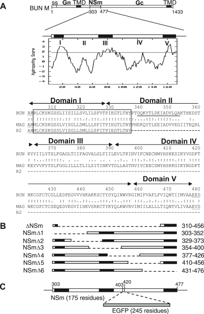
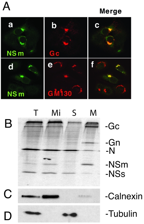
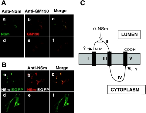
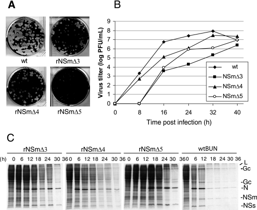
The nonstructural protein NSm of Bunyamwera virus (BUNV), the prototype of the Bunyaviridae family, is encoded by the M segment in a polyprotein precursor, along with the virion glycoproteins, in the order Gn-NSm-Gc. As little is known of its function, we examined the intracellular localization, membrane integrality, and topology of NSm and its role in virus replication. We confirmed that NSm is an integral membrane protein and that it localizes in the Golgi complex, together with Gn and Gc. Coimmunoprecipitation assays and yeast two-hybrid analysis demonstrated that NSm was able to interact with other viral proteins. NSm is predicted to contain three hydrophobic (I, III, and V) and two nonhydrophobic (II and IV) domains. The N-terminal nonhydrophobic domain II was found in the lumen of an intracellular compartment. A novel BUNV assembly assay was developed to monitor the formation of infectious virus-like-particles (VLPs). Using this assay, we showed that deletions of either the complete NSm coding region or domains I, II, and V individually seriously compromised VLP production. Consistently, we were unable to rescue viable viruses by reverse genetics from cDNA constructs that contained the same deletions. However, we could generate mutant BUNV with deletions in NSm domains III and IV and also a recombinant virus with the green fluorescent protein open reading frame inserted into NSm domain IV. The mutant viruses displayed differences in their growth properties. Overall, our data showed that the N-terminal region of NSm, which includes domain I and part of domain II, is required for virus assembly and that the C-terminal hydrophobic domain V may function as an internal signal sequence for the Gc glycoprotein.


Figures

FIG. 1.
FIG. 1.
The BUNV M RNA segment and the encoded precursor polyprotein. (A) The gene layout of the M segment is shown at the top, with positions of amino acid residues marking protein boundaries indicated. ss, signal peptide; TMD, transmembrane domain. Below, the Kyte-Doolittle hydropathy plot and predicted domain structure of NSm are shown. Domains I to V were suggested by the program TMHMM (21). The amino acid alignment of the NSm proteins of BUNV, Maguari virus, and its mutant R2 are shown, with the conserved N-terminal region boxed. The peptide sequence used to raise the anti-NSm antibody is underlined. (B) Schematic of NSm deletion mutants. The regions deleted are indicated by the dashed line, and the residues deleted are indicated at the right. (C) Insertion of EGFP open reading frame into NSm. The EGFP coding sequence was cloned into the M segment cDNA at artificial SacI restriction enzyme sites created at codons 403 and 420 in NSm.
FIG. 2.
FIG. 2.
Intracellular localization and determination of the membrane integrality of NSm. (A) Colocalization of NSm with Gc (panels a to c) and Golgi matrix protein GM130 (panels d to f). wt BUNV-infected BSR-T7/5 cells were stained with a mixture of anti-NSm antibody and either anti-Gc MAb 742 or anti-GM130 MAb. NSm stains green (panels a and d), and Gc and GM130 stain red. Merged confocal microscopic images are also shown, with colocalization shown in yellow (panels c and f). (B) NSm is an integral membrane protein. Vero E6 cells were infected with wt BUNV and radiolabeled with [35S]methionine, and membrane fractions were prepared as described in Materials and Methods. Total (T) and microsomal (Mi) fractions were collected, and membranes were extracted with sodium carbonate to yield supernatant (S) and membrane (M) samples. The fractions were analyzed by SDS-PAGE. The positions of viral proteins are indicated at the right. (C). Western blot analysis of the gel using anti-calnexin antibodies as a marker for membranes. (D) Western blot analysis of the gel using anti-tubulin antibodies as a marker for the cytosolic fraction.
FIG. 3.
FIG. 3.
Determination of the topology of BUNV NSm. Vero E6 cells were infected with wt BUNV (A) or recombinant virus rBUNM-NSm-EGFP (B) and semipermeabilized by the freeze-thaw technique. Cells shown in the upper row of each set were further permeabilized with 0.2% Triton X-100-PBS (panels a to c in each case). Before examination by confocal microscopy, the wt BUNV-infected cells were costained with rabbit anti-NSm serum and anti-GM130 MAb and the rBUNM-NSm-EGFP-infected cells were stained only with anti-NSm serum. In panel A, NSm stains green and GM130 stains red, and in panel B, the EGFP autofluorescence of NSm-EGFP protein shows as green and the NSm antibody stains red. Merged confocal images are also shown. NSm antibodies can only react with NSm or NSm-EGFP in fully permeabilized cells. (C) The predicted topology of NSm. Hydrophobic domains are shown as black columns across the intracellular membrane. The positions of the predicted domains I to V and of the anti-NSm epitope are marked.
FIG. 4.
FIG. 4.
Effect of internal deletions in NSm on virus assembly, protein processing, and intracellular transport. (A) Production of virus-like particles. BSR-T7/5 cells were transfected with minigenome-component plasmids and either wt BUNV M segment cDNA or mutated NSm-containing plasmids as indicated. Controls included pTM1 instead of pTM1-BUNM, substitution of wt BUNV L segment cDNA with an inactive L cDNA mutant, and mock-infected cells. Supernatants from these cells were taken 24 h posttransfection and used to infect fresh BSR-T7/5 cells that had been transfected with BUNV L and N protein-expressing plasmids 5 h previously. In one case, the supernatant was reacted with anti-BUN antibody prior to infection. Renilla luciferase activity in all extracts of these cells was measured after 24 h and is shown in arbitrary light units. 1, pTM1 vector control; 2, pTM1-BUNM (wt control); 3, anti-BUNV serum-neutralized supernatant from pTM1-BUN-M-transfected cells; 4, pTM1-BUNM-NSmΔ1; 5, pTM1-BUNM-NSmΔ2; 6, pTM1-BUNM-NSmΔ3; 7, pTM1-BUNM-NSmΔ4; 8, pTM1-BUNM-NSmΔ5; 9, pTM1-BUNM-NSmΔ6; 10, pTM1-BUNMΔNSm; 11, substitution with inactive L mutant; 12, mock-transfected cells. (B) Processing of BUNV glycoproteins from Vero E6 cells transfected with wt or NSm deletion mutant cDNA clones. Vero E6 cells were infected with recombinant vaccinia virus vTF7-3, followed by transfection with pTM1-BUNM (wt) or NSm mutant cDNA constructs. Cells were labeled with [35S]methionine for 15 h, extracts were prepared, and viral proteins were immunoprecipitated with anti-BUN serum. The labeled glycoproteins were subjected to endo H (H) or mock digestion (C) and analyzed by SDS-10% PAGE under reducing conditions. The relevant portions of the gels are shown. (C) Intracellular localization of Gc expressed from wt and mutated NSm-containing M segment cDNAs. BSR-T7/5 cells were transfected cDNA constructs as indicated and were stained with a mixture of anti-Gc MAb 742 and anti-GM130 antibodies and 4′,6′-diamidino-2-phenylindole (DAPI). Cells were examined using the DeltaVision microscopy system (AppliedPrecision), and triple-stained images are shown. Gc proteins stain red, the Golgi stains green, and cell nuclei, stained with DAPI, are shown in blue.
FIG. 5.
FIG. 5.
Protein profiles of cells infected with wild-type and recombinant BUNV. Vero E6 cells were infected with wt BUNV (lane 2), rBUNM-NSmΔ3 (lane 3), rBUNM-NSmΔ4 (lane 4), rBUNM-NSmΔ5 (lane 5), and rBUNM-NSm-EGFP (lane 6) at 5 PFU/cell. At 24 hpi, cells were labeled with 100 μCi [35S]methionine for 2 h, and then equal amounts of cell lysate were analyzed by SDS-12.5% PAGE under reducing conditions. Positions of viral proteins are indicated.
FIG. 6.
FIG. 6.
Plaque phenotype, growth kinetics, and protein synthesis shutoff of wt and mutant BUNV. (A) Comparison of plaque morphology on Vero E6 cells. Cell monolayers were fixed with 4% formaldehyde and stained with Giemsa solution 4 days after infection. (B) Viral growth curves. Vero E6 cells were infected with either wt or recombinant viruses at an MOI of 0.01 PFU/cell. Virus was harvested at 8-h intervals and titrated by plaque assay. The results shown are the averages from two independent titrations. (C) Time course of protein synthesis. Vero E6 cells infected at an MOI of 1.0 PFU/cell were labeled with 100 μCi [35S]methionine for 20 min at the time points indicated, and cell lysates were analyzed by SDS-15% PAGE. The positions of the viral proteins are indicated at the right.
FIG. 7.
FIG. 7.
Interaction of Bunyamwera virus proteins. (A) Coimmunoprecipitation. BUNV-infected Vero E6 cells (at 5 PFU/cell) were labeled with 80 μCi [35S]methionine for 4 h at 30 h postinfection, and equal volumes of cell lysate were immunoprecipitated with anti (α)-BUN serum (lanes 1 and 2), anti-Gc MAb 742 (lanes 3 and 4), anti-NSm serum (lanes 5 and 6), or anti-N serum (lanes 7 and 8). I, BUNV-infected cells; C, mock-infected control cells. The positions of the viral proteins are indicated. (B) Interaction studied by yeast two-hybrid analysis. S. cerevisiae AH109 cells were cotransformed with plasmids as listed below and plated on selective medium as described in Materials and Methods. Plasmid combinations: a, pBK-NSm + pAD-NSm; b, pBK + pAD-NSm; c, pBK-NSm + pAD; d, pBK-NSs + pAD-NSm; e, pBK-NSs + pAD; f, negative control; g, pBK-T + pAD-p53; h, pBK-T + pAD-Lam; i, pBK-Gn + pAD; j, pBK-Gn + pAD-NSm; k, pBK + pAD-NSm; l, negative control. Growth on the plates indicates interaction of NSm with itself (sector a), with NSs (sector d), and the cytoplasmic tail of Gn (sector j).

Similar articles

Cited by

References

    1. Bishop, D. H. 1996. Biology and molecular biology of bunyaviruses, p. 19-61. In R. M. Elliott (ed.), The Bunyaviridae. Plenum Press, New York, N.Y.
    1. Bridgen, A., and R. M. Elliott. 1996. Rescue of a segmented negative-strand RNA virus entirely from cloned complementary DNAs. Proc. Natl. Acad. Sci. USA 93:15400-15404. - PMC - PubMed
    1. Bridgen, A., F. Weber, J. K. Fazakerley, and R. M. Elliott. 2001. Bunyamwera bunyavirus nonstructural protein NSs is a nonessential gene product that contributes to viral pathogenesis. Proc. Natl. Acad. Sci. USA 98:664-669. - PMC - PubMed
    1. Buchholz, U. J., S. Finke, and K. K. Conzelmann. 1999. Generation of bovine respiratory syncytial virus (BRSV) from cDNA: BRSV NS2 is not essential for virus replication in tissue culture, and the human RSV leader region acts as a functional BRSV genome promoter. J. Virol. 73:251-259. - PMC - PubMed
    1. Bupp, K., K. Stillmock, and F. Gonzalez-Scarano. 1996. Analysis of the intracellular transport properties of recombinant La Crosse virus glycoproteins. Virology 220:485-490. - PubMed

Publication types

Substances