Compensatory substitutions restore normal core assembly in simian immunodeficiency virus isolates with Gag epitope cytotoxic T-lymphocyte escape mutations
- PMID: 16873273
- PMCID: PMC1563819
- DOI: 10.1128/JVI.00068-06
Compensatory substitutions restore normal core assembly in simian immunodeficiency virus isolates with Gag epitope cytotoxic T-lymphocyte escape mutations
Abstract
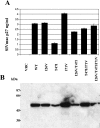
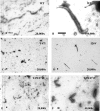
The evolution of human immunodeficiency virus type 1 (HIV-1) and simian immunodeficiency virus (SIV) as they replicate in infected individuals reflects a balance between the pressure on the virus to mutate away from recognition by dominant epitope-specific cytotoxic T lymphocytes (CTL) and the structural constraints on the virus' ability to mutate. To gain a further understanding of the strategies employed by these viruses to maintain replication competency in the face of the intense selection pressure exerted by CTL, we have examined the replication fitness and morphological ramifications of a dominant epitope mutation and associated flanking amino acid substitutions on the capsid protein (CA) of SIV/simian-human immunodeficiency virus (SHIV). We show that a residue 2 mutation in the immunodominant p11C, C-M epitope (T47I) of SIV/SHIV not only decreased CA protein expression and viral replication, but it also blocked CA assembly in vitro and virion core condensation in vivo. However, these defects were restored by the introduction of upstream I26V and/or downstream I71V substitutions in CA. These findings demonstrate how flanking compensatory amino acid substitutions can facilitate viral escape from a dominant epitope-specific CTL response through the effects of these associated mutations on the structural integrity of SIV/SHIV.
Figures


Similar articles
-
Simian-human immunodeficiency virus escape from cytotoxic T-lymphocyte recognition at a structurally constrained epitope.J Virol. 2003 Dec;77(23):12572-8. doi: 10.1128/jvi.77.23.12572-12578.2003. J Virol. 2003. PMID: 14610180 Free PMC article.
-
Fitness costs limit viral escape from cytotoxic T lymphocytes at a structurally constrained epitope.J Virol. 2004 Dec;78(24):13901-10. doi: 10.1128/JVI.78.24.13901-13910.2004. J Virol. 2004. PMID: 15564498 Free PMC article.
-
Cytotoxic T lymphocytes do not appear to select for mutations in an immunodominant epitope of simian immunodeficiency virus gag.J Immunol. 1992 Dec 15;149(12):4060-6. J Immunol. 1992. PMID: 1460291
-
Structural constraints on viral escape from HIV- and SIV-specific cytotoxic T-lymphocytes.Viral Immunol. 2004;17(2):144-51. doi: 10.1089/0882824041310658. Viral Immunol. 2004. PMID: 15279695 Review.
-
Genetic and biochemical studies on the assembly of an enveloped virus.Genet Eng (N Y). 2001;23:83-112. doi: 10.1007/0-306-47572-3_6. Genet Eng (N Y). 2001. PMID: 11570108 Review. No abstract available.
Cited by
-
Phylogenetic dependency networks: inferring patterns of CTL escape and codon covariation in HIV-1 Gag.PLoS Comput Biol. 2008 Nov;4(11):e1000225. doi: 10.1371/journal.pcbi.1000225. Epub 2008 Nov 21. PLoS Comput Biol. 2008. PMID: 19023406 Free PMC article.
-
Recovery of fitness of a live attenuated simian immunodeficiency virus through compensation in both the coding and non-coding regions of the viral genome.Retrovirology. 2007 Jul 3;4:44. doi: 10.1186/1742-4690-4-44. Retrovirology. 2007. PMID: 17608929 Free PMC article.
-
Molecular evolution of human immunodeficiency virus type 1 upon transmission between human leukocyte antigen disparate donor-recipient pairs.PLoS One. 2008 Jun 18;3(6):e2422. doi: 10.1371/journal.pone.0002422. PLoS One. 2008. PMID: 18560583 Free PMC article.
-
Composite Sequence-Structure Stability Models as Screening Tools for Identifying Vulnerable Targets for HIV Drug and Vaccine Development.Viruses. 2015 Nov 4;7(11):5718-35. doi: 10.3390/v7112901. Viruses. 2015. PMID: 26556362 Free PMC article.
-
HLA class I-driven evolution of human immunodeficiency virus type 1 subtype c proteome: immune escape and viral load.J Virol. 2008 Jul;82(13):6434-46. doi: 10.1128/JVI.02455-07. Epub 2008 Apr 23. J Virol. 2008. PMID: 18434400 Free PMC article.
References
-
- Allen, T. M., D. H. O'Connor, P. Jing, J. L. Dzuris, B. R. Mothe, T. U. Vogel, E. Dunphy, M. E. Liebl, C. Emerson, N. Wilson, K. J. Kunstman, X. Wang, D. B. Allison, A. L. Hughes, R. C. Desrosiers, J. D. Altman, S. M. Wolinsky, A. Sette, and D. I. Watkins. 2000. Tat-specific cytotoxic T lymphocytes select for SIV escape variants during resolution of primary viraemia. Nature 407:386-390. - PubMed
-
- Barouch, D. H., J. Kunstman, J. Glowczwskie, K. J. Kunstman, M. A. Egan, F. W. Peyerl, S. Santra, M. J. Kuroda, J. E. Schmitz, K. Beaudry, G. R. Krivulka, M. A. Lifton, D. A. Gorgone, S. M. Wolinsky, and N. L. Letvin. 2003. Viral escape from dominant simian immunodeficiency virus epitope-specific cytotoxic T lymphocytes in DNA-vaccinated rhesus monkeys. J. Virol. 77:7367-7375. - PMC - PubMed
-
- Barouch, D. H., J. Kunstman, M. J. Kuroda, J. E. Schmitz, S. Santra, F. W. Peyerl, G. R. Krivulka, K. Beaudry, M. A. Lifton, D. A. Gorgone, D. C. Montefiori, M. G. Lewis, S. M. Wolinsky, and N. L. Letvin. 2002. Eventual AIDS vaccine failure in a rhesus monkey by viral escape from cytotoxic T lymphocytes. Nature 415:335-339. - PubMed
-
- Borrow, P., H. Lewicki, X. Wei, M. S. Horwitz, N. Peffer, H. Meyers, J. A. Nelson, J. E. Gairin, B. H. Hahn, M. B. Oldstone, and G. M. Shaw. 1997. Antiviral pressure exerted by HIV-1-specific cytotoxic T lymphocytes (CTLs) during primary infection demonstrated by rapid selection of CTL escape virus. Nat. Med. 3:205-211. - PubMed
Publication types
MeSH terms
Substances
Grants and funding
LinkOut - more resources
Full Text Sources

