. 2006 Oct 15;576(Pt 2):569-83.
doi: 10.1113/jphysiol.2006.115766. Epub 2006 Jul 27.

A spinal vasopressinergic mechanism mediates hyperosmolality-induced sympathoexcitation

Affiliations

A spinal vasopressinergic mechanism mediates hyperosmolality-induced sympathoexcitation

V R Antunes et al. J Physiol. .

Abstract

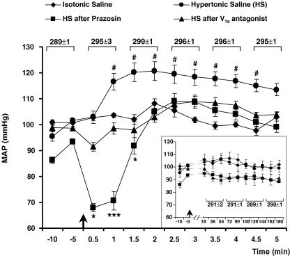
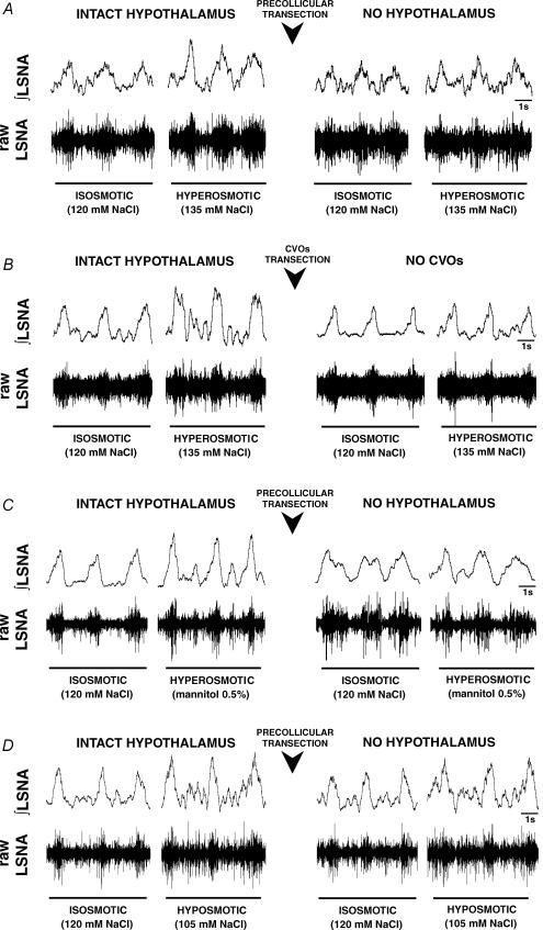
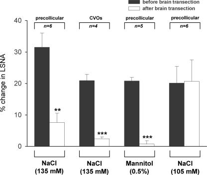
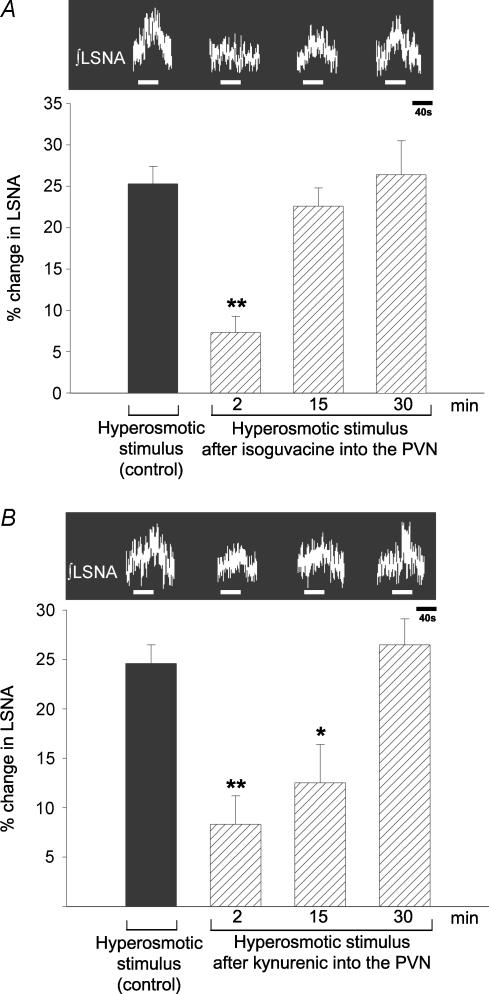
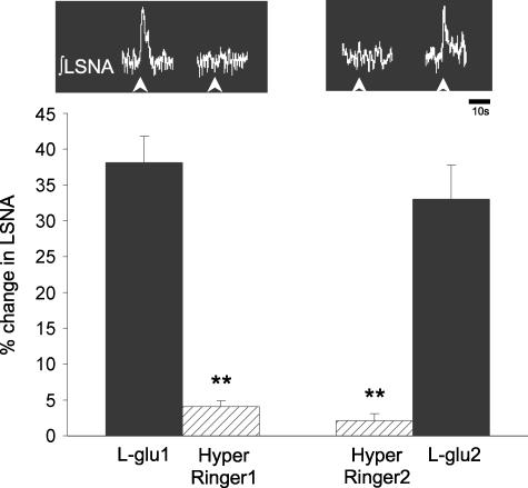
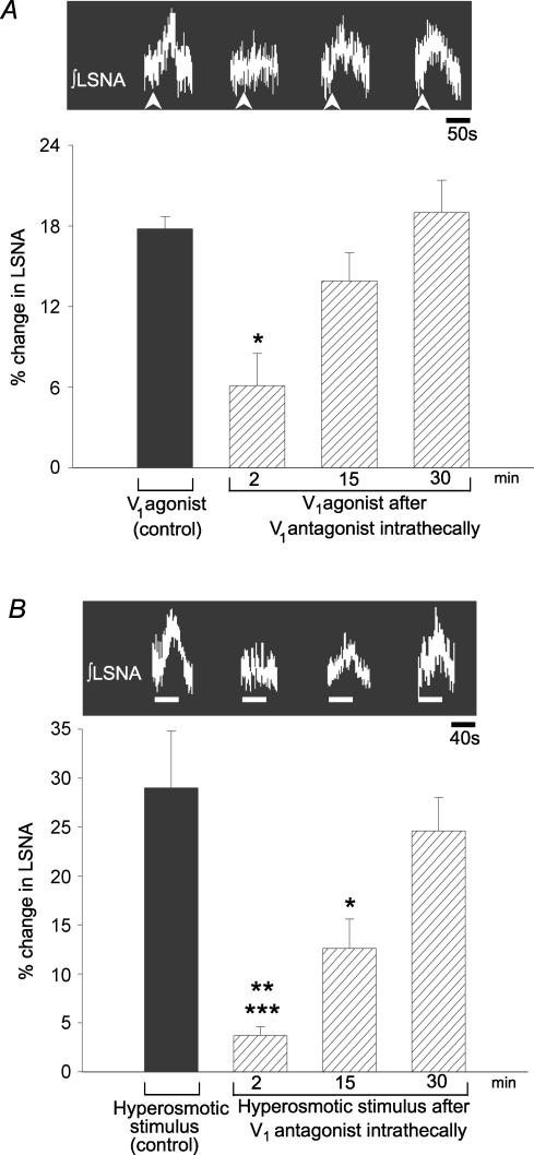
An elevation in plasma osmolality elicits a complex neurohumoral response, including an activation of the sympathetic nervous system and an increase in arterial pressure. Using a combination of in vivo and in situ rat preparations, we sought to investigate whether hypothalamic vasopressinergic spinally projecting neurones are activated during increases in plasma osmolality to elicit sympathoexcitation. Hypertonic saline (HS, i.v. bolus), which produced a physiological increase in plasma osmolality to 299 +/- 1 mosmol (kg water)(-1), elicited an immediate increase in mean arterial pressure (MAP) (from 101 +/- 1 to 121 +/- 3 mmHg) in vivo. Pre-treatment with prazosin reversed the HS-induced pressor response to a hypotensive response (from 121 +/- 3 to 68 +/- 2 mmHg), indicating significant activation of the sympathetic nervous system. In an in situ arterially perfused decorticate rat preparation, hyperosmotic perfusate consisted of either 135 mm NaCl, or a non-NaCl osmolyte, mannitol (0.5%); both increased lumbar sympathetic nerve activity (LSNA) by 32 +/- 5% (NaCl) and 21 +/- 1% (mannitol), which was attenuated after precollicular transection (7 +/- 3% and 1 +/- 1%, respectively). Remaining experiments used the NaCl hyperosmotic stimulus. In separate preparations the hyperosmotic-induced sympathoexcitation (21 +/- 2%) was also significantly attenuated after transection of the circumventricular organs (2 +/- 1%). Either isoguvacine (a GABA(A) receptor agonist) or kynurenic acid (a non-selective ionotropic glutamate receptor antagonist) microinjected bilaterally into the paraventricular nucleus (PVN) attenuated the increase in LSNA induced by the hyperosmotic stimulus (control: 25 +/- 2%; after isoguvacine: 7 +/- 2%; after kynurenic: 8 +/- 3%). Intrathecal injection of a V(1a) receptor antagonist also reduced the increase in LSNA elicited by the hyperosmotic stimulus (control: 29 +/- 6%; after blocker: 4 +/- 1%). These results suggest that a physiological hyperosmotic stimulus produces sympathetically mediated hypertension in conscious rats. These data are substantiated by the in situ decorticate preparation in which sympathoexcitation was also evoked by comparable hyperosmotic stimulation. Our findings demonstrate the importance of vasopressin acting on spinal V(1a) receptors for mediating sympathoexcitatory response to acute salt loading.


Figures

Figure 7
Figure 7
A, photomicrographs of a sagittal brain section, taken 0.1 mm lateral to the midline from a decorticated in situ preparation showing the intact lamina terminalis. The magnified areas depict the SFO and MnPO at higher magnification, respectively. The arrowheads indicate the level of the transection to disconnect the CVOs from the PVN. B, photomicrographs of a sagittal section, taken from the same brain as shown in A, 0.3 mm lateral to the midline. The magnified area shows that the transection (indicated by arrowheads) was made rostral to the PVN. The arrowheads indicate the level of transection. Scale bars: 200 μm. C, representative photomicrograph of the site of a bilateral microinjection into the PVN. The arrows mark the position of the glass micropipette tip. D, schematic diagram of coronal sections of the hypothalamus at the level of PVN modified from the atlas of Paxinos & Watson (1996), showing the centre of bilateral microinjections of isoguvacine (^), kynurenic acid (□) or unilateral microinjections of l-glutamate and hyperosmotic Ringer solution (▵) into the PVN. Asterisks (*) represent microinjection sites of isoguvacine and kynurenic acid outside of the PVN, in a group of three rats that were ineffective in altering HS-induced sympathoexcitatory responses. Abbreviations: AC, anterior commissure; SFO, subfornical organ; MnPO, median preoptic nucleus; PVN, paraventricular hypothalamic nucleus; PaDC, paraventricular hypothalamic nucleus, dorsal cap; PaLM, paraventricular hypothalamic nucleus, lateral magnocellular part; PaMP, paraventricular hypothalamic nucleus, medial parvicellular part; PaV, paraventricular hypothalamic nucleus, ventral part; Pe, periventricular hypothalamic nucleus; ME, median eminence; Arc, arcuate hypothalamic nucleus; 3V, third ventricle.
Figure 1
Figure 1. Intravenous injections of hyperosmotic saline (HS) increase arterial pressure in conscious rats
HS (3 m, i.v.) produced an increase in plasma osmolality (values show are per min for 5 min) and in the MAP, which reached a significant peak at 90 s (299 ± 1 mosmol (kg water)−1). Prazosin reversed the pressor response evoked by HS into a depressor effect at 30 and 60 s (n =5). HS injection failed to produce an increase in the MAP after administration of a V1a receptor antagonist. Isotonic saline injected intravenously produced a negligible effect on the MAP (n =5). Inset graph, the pressor response evoked by HS recovered after 180 min and plasma osmolality values are show every 40 min. Arrows show the time when HS or isotonic saline were injected. #P < 0.05 compared to isotonic saline injection. *P < 0.05 and ***P < 0.001 compared to HS injection over the same time course. Error bars show s.e.m.
Figure 2
Figure 2. Lumbar sympathetic nerve activity (LSNA) changes during three distinct osmotic conditions (isosmotic, hyperosmotic and hyposmotic) before and after precollicular transection and selective ablation of the CVOs
Representative traces of changes in raw and integrated (∫) LSNA during isosmotic (120 mm NaCl) and hyperosmotic stimuli (135 mm NaCl) before (intact hypothalamus) and after precollicular brain transection (no hypothalamus, A) or CVOs transection (B). Recordings in C show the sympathoexcitatory effect of mannitol (0.5%) before and after precollicular transection. A hyposmotic stimulus (105 mm NaCl, D) also produced sympathoexcitation, but this was not affected by precollicular brain transection. **P < 0.01 and ***P < 0.001 compared to before brain transection.
Figure 3
Figure 3. LSNA before and after brain transection (precollicular or CVOs) induced by hyperosmotic (135 mM NaCl or mannitol 0.5%) or hyposmotic (105 mM NaCl) stimuli in the in situ rat preparation
n is the number of preparations utilized in every protocol. **P < 0.01 and ***P < 0.001 compared to before brain transection. Error bars are s.e.m.
Figure 4
Figure 4. Reversible inactivation of the PVN and blockade of excitatory amino acid (EAA) receptors within the PVN reduce hyperosmotic-induced sympathoexcitation in situ
A, isoguvacine (1 mm; 60 nl) microinjected bilaterally into the PVN attenuated the increase in the LSNA elicited by the hyperosmotic stimulus. This effect reversed 15 and 30 min later. **P < 0.01 compared to control, 15 and 30 min; (n =5). B, blockade of EAA receptors with kynurenic acid (100 mm, 60 nl) microinjected bilaterally into the PVN attenuated the increase in LSNA elicited by a hyperosmotic stimulus; total recovery was at 30 min **P < 0.01 and *P < 0.05 compared to control and 30 min, respectively; (n =5). Hatched bars: hyperosmotic stimulus (40 s) after microinjections into the PVN. Error bars are s.e.m.
Figure 5
Figure 5. Effect of hyperosmotic perfusate microinjected directly into the PVN on the lumbar sympathetic nerve activity in situ
L-Glutamate (L-glu, 20 mM) or hyperosmotic Ringer solution (Hyper Ringer solution, 320 mosmol (kg water)−1) was microinjected unilaterally into the PVN. Despite switching the order in which the different injections were made, hyperosmotic injections were ineffective. In each case there was a 30-min interval between each microinjection. **P < 0.01 compared to l-glu1 and l-glu2; (n =3). Arrowheads show the time when l-glutamate or hyperosmotic Ringer solution were microinjected into the PVN. Error bars are s.e.m.
Figure 6
Figure 6. Intrathecal injection of a V1a receptor blocker reduces the hyperosmotically induced sympathoexcitation in situ
A, the efficacy of a V1a receptor antagonist (1 μg ml−1) was tested by assessing whether it attenuated the increases in LSNA elicited by V1a receptor agonist (1 μg ml−1) via intrathecal injections. *P < 0.05 compared to control; (n =3). Arrowheads show the time when V1a agonist was microinjected into the PVN. B, intrathecal injection of the V1a receptor antagonist (1 μg ml−1) attenuated the increases in the LSNA elicited by hyperosmotic stimulus (control) at 2 and 15 min. The response recovered 30 min later. ***P < 0.001 and *P < 0.05 compared to control; **P < 0.01, compared to 30 min; (n =5). Error bars are s.e.m.

Similar articles

Cited by

References

    1. Akins VF, Bealer SL. Brain histamine regulates pressor responses to peripheral hyperosmolality. Am J Physiol. 1990;259:R507–R513. - PubMed
    1. Antunes VR, Yao ST, Murphy D, Pickering AE, Paton JFR. An in situ preparation for studying hypothalamic control of sympathetic activity. J Physiol. 2005;567P:PC51.
    1. Antunes-Rodrigues J, de Castro M, Elias LL, Valenca MM, McCann SM. Neuroendocrine control of body fluid metabolism. Physiol Rev. 2004;84:169–208. - PubMed
    1. Badoer E. Cardiovascular role of parvocellular neurones in the paraventricular nucleus of the hypothalamus. News Physiol Sci. 1996;11:43–47.
    1. Baertschi AJ, Vallet PG. Osmosensitivity of the hepatic portal vein area and vasopressin release in rats. J Physiol. 1981;315:217–230. - PMC - PubMed

Publication types

MeSH terms

LinkOut - more resources