. 2006 Oct 1;576(Pt 1):309-27.
doi: 10.1113/jphysiol.2006.110577. Epub 2006 Jul 27.

Local and diffuse mechanisms of primary afferent depolarization and presynaptic inhibition in the rat spinal cord

Affiliations

Local and diffuse mechanisms of primary afferent depolarization and presynaptic inhibition in the rat spinal cord

Malcolm Lidierth. J Physiol. .

Abstract

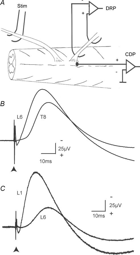
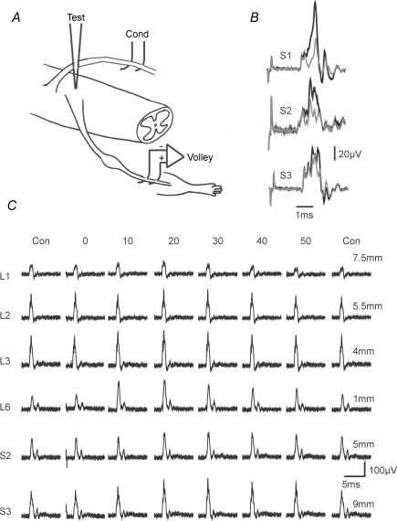
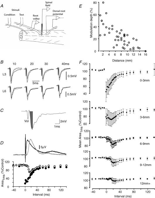
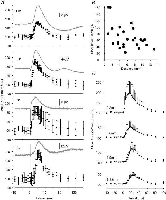
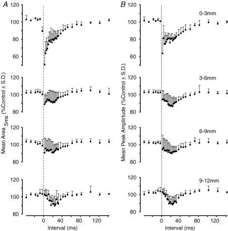
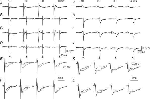
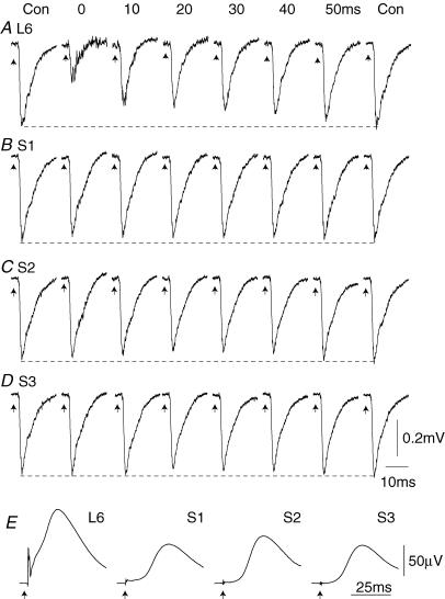
Two types of dorsal root potential (DRP) were found in the spinal cord of urethane-anaesthetized rats. Local DRPs with short latency-to-onset were evoked on roots close to the point of entry of an afferent volley. Diffuse DRPs with a longer latency-to-onset were seen on more distant roots up to 17 segments from the volley entry zone. The switch to long latency-to-onset occurred abruptly as a function of distance along the cord and could not be explained by conduction delays within the dorsal columns. Long-latency DRPs were also present and superimposed on the short-latency DRPs on nearby roots. Both local and diffuse DRPs were evoked by light mechanical stimuli: von Frey hair thresholds were <or= 1 gram force Changes in excitability of the terminals of sural nerve afferents were used to confirm that both local and diffuse DRPs were associated with primary afferent depolarization (PAD). These effects were potent: the area of the antidromic volley evoked in the sural nerve by intraspinal microstimulation in the L4/5 spinal segment was increased by 109 +/- 50% (mean +/- s.d.; n = 5) by nearby conditioning stimuli, and by 52 +/- 12% (n = 6) with stimuli applied 9-13 mm (5-8 segments) away. The time course of the changes in terminal excitability closely matched those of the DRPs. Reduction of the field potentials evoked in the dorsal horn by stimulation of dorsal roots was also shown to accompany both local and diffuse DRPs. The area of the monosynaptically evoked field potential was reduced by 48 +/- 19% (n = 7) with nearby conditioning stimulation and 16 +/- 9% (n = 10) with stimulation 9-12 mm distant. Evidence is presented that this inhibition includes a presynaptic component. Similar effects were seen with field potentials evoked by sural nerve stimulation. It is concluded that diffuse DRPs are mediated through propriospinal networks which may contribute to the gating of sensory information flow during natural behaviour as they respond to weak mechanical stimuli and provoke presynaptic inhibition.


Figures

Figure 1
Figure 1. Near and distant DRPs
A, the experimental arrangement for stimulating dorsal roots and recording the DRP and CDP. By convention (Barron & Matthews, 1938), the proximal electrode is connected to the inverting input of the amplifier and DRPs are illustrated negative-up. Typical DRPs are shown in B and C. B, a stimulus to the L5 root (10 μA; 200 μs pulse) evoked a DRP on the nearby L6 dorsal root (2 mm) and also on the more distant T8 root located 17 mm away. The arrowhead marks the time of the stimulus. The spinal cord had been transected at mid-thoracic level in this animal. C, similar potentials from a preparation with an intact spinal cord. The stimulus was to the T13 dorsal root and recordings were made at L1 (distance 2.5 mm) and L6 (11.5 mm).
Figure 5
Figure 5. Changes in excitability of sural nerve terminals
A, the experimental arrangement. B, specimen averaged antidromic volleys recorded from the sural nerve. The effects of conditioning stimuli of 100 μA strength were examined when applied to the S1, S2 or S3 roots. Test stimuli were of 6.0, 8.4 and 6.0 μA, respectively, and were delivered to the dorsal horn in the L5 segment (for conditioning stimulation of S1 and S3 roots) or at the L4/5 boundary (S2) at interstimulus intervals of 14 ms (S1) or 22 ms (S2 and 3). Control (grey) and test (black) responses are superimposed. C, the time course of this effect with stimulation of roots located rostral to the test stimulus (L1, distance 7.5 mm; L2, 5.5 mm and L3, 4 mm) and caudal (L6, 1 mm; S2, 5 mm and S3, 9 mm). Data are from a single experiment.
Figure 7
Figure 7. Effects of conditioning stimuli on dorsal root-evoked field potentials in the dorsal horn
A, the experimental setup. B, the responses recorded at depth in the dorsal horn of L1 following electrical stimulation of the L1 dorsal root. Control traces are shown (grey lines), together with traces (black lines) that were conditioned by preceding stimulation of the dorsal roots of L3 (upper traces; 4 mm distant) and L6 (lower traces; 10 mm distant) at intervals of 10–40 ms as indicated. C shows how the monosynaptic component of the evoked field potential was estimated. The grey filled area (Area1ms) covers the negative component of the field up to 1 ms after the peak of the afferent volley recorded at depth (‘Vol’). D, the full time course of the effects of conditioning stimulation on this component of the evoked field potential. Field potentials were recorded in the L2 dorsal horn and evoked by L2 dorsal root stimulation. Conditioning stimuli were delivered to the dorsal roots of L3 (black circles) or L6 (grey circles) segments and are shown together with the DRPs evoked on the L1 root. Data are from a different preparation to B. E, the modulation depth measured from graphs such as those in D, as a function of distance. Data are from 42 root pairs in 12 animals. F, averaged time courses for the inhibition of the monosynaptic component of dorsal horn field potentials calculated from pooled data. Test stimuli (range 0.7–10 μA) were delivered to the dorsal root of the recorded segment and conditioned by stimuli (10 or 100 μA) to dorsal roots with entry zones located, from above downwards, 0–3 mm from the recording point (n = 7), 3–6 mm (n = 7), 6–9 mm (n = 11), 9–12 mm (n = 10) and more than 12 mm (n = 7). Error bars show standard deviations. Data were derived from 12 rats.
Figure 6
Figure 6. Magnitude and time course of the changes in excitability of sural nerve terminals
A, a quantitative analysis of the data from an experiment in which conditioning stimuli were applied to the T12, L2, S1 or S2 dorsal roots (distances from the test-stimulus site of 9.5, 4, 2 and 7 mm, respectively). Test stimuli in the L5 segment were 4.3–6.4 μA. Conditioning stimuli were 100 μA in all cases. The DRPs recorded subsequently with stimulation of each conditioning root are superimposed on the graphs. B and C, summarize the data from experiments where graphs such as those shown in A were constructed with conditioning stimulation of 26 roots at various distances from the test-stimulation site. B, the depth of modulation (see Methods) as a function of distance for each of the 26 roots. C, data were pooled to allow an average time course to be calculated for samples where the conditioning root was 0–3 mm distant (n = 5), 3–6 mm (n = 9), 6–9 mm (n = 6) or 9–13 mm (n = 6).
Figure 10
Figure 10. Averaged effects of conditioning stimuli on sural nerve-evoked field potentials in the dorsal horn
In A and B, the effects of conditioning stimuli for a range of interstimulus intervals, and a range of intersegmental distances, are quantified. A, the change in area of the first 5 ms of the evoked field. B, the change in peak amplitude. Data were pooled from 11 preparations (0–3 mm, n = 3 trials; 3–6 mm, n = 14; 6–9 mm, n = 8; 9–12 mm, n = 5). Interstimulus intervals on the abscissa have been corrected for the additional conduction delay in the sural nerve (2.2 ± 0.13 ms, mean ± s.d., n = 11).
Figure 2
Figure 2. Effect of distance on the latency and form of the DRP
A, averaged DRPs evoked by a stimulus to the third coccygeal dorsal root (Co3) and recorded on successively more rostral dorsal roots, or divided rootlets, up to the T10 spinal level in a single animal. Each trace has been positioned so that the vertical displacement of the prestimulus period is proportional to distance along the cord (5 mm calibration bar shown; note that the voltage calibration is different for the upper 3 traces). B, as in A, but from another preparation in which the T11 dorsal root was stimulated and DRPs were recorded from successively more caudal roots. C and D, averaged DRPs (black lines) and CDPs (grey lines) evoked at the L3 level in response to stimulation of more caudal dorsal roots (L4, 0.5 mm separation; L6 5.5 mm and S3, 11 mm). The traces are shown on a slow time base in C and a fast time base in D. Stimuli were 10 μA, 200 μs pulses in all cases. Traces in C and D are from a single animal.
Figure 3
Figure 3. Comparison of mechanically and electrically evoked DRPs
A, the DRPs evoked at L5 and T12 levels by five successive taps to the plantar surface of the ipsilateral hindpaw with a blunt probe. B, shows the averaged response to the paw tap (left; 22 stimuli) together with that evoked by an electrical stimulus to the L6 dorsal root (right; 10 μA, 200 μs stimulus, 43 stimuli). The traces in B have been scaled to equal heights to assist comparison of the timecourses. Data are from a single preparation. In C, the delays between the peaks of the DRP on thoracic roots (T9–T12) and those on L5 roots are plotted for 10 animals. The line in C is the line of equality.
Figure 4
Figure 4. The effects of picrotoxin on the amplitude of the DRP
A, the data from a single experiment in which the L6 dorsal root was stimulated at 100 × T while recording the DRPs at the L5 (•) and T11 (○) levels. DRPs were averaged over 30 stimuli and their amplitudes are plotted as a function of time. Picrotoxin was administered intravenously at the times indicated. Doses are cumulative. The insets show specimen averaged traces for each dose together with a control trace. B, the pooled results from five animals (three animals where both L5 and T11 DRPs were successfully recorded over the course of the experiment and two others in which only one of the L5 or T11 DRPs was recorded throughout). Data shown are means ± s.d. (n = 4 for doses of 2–50 mg kg−1, n = 3 for 100 mg kg−1).
Figure 8
Figure 8. Specimen traces showing the effects of conditioning stimulation on the responses evoked in the dorsal horn of the L2 segment
Test stimuli were delivered to the L2 dorsal root. In A–F, conditioning stimuli were delivered to the L3 dorsal root (2 mm distant from the recording site) while in G–L, conditioning stimuli were delivered to the L6 root (8 mm distant). A and G, the effects of conditioning stimuli delivered alone. B and H, the control responses. C and I, the test responses evoked following conditioning stimulation 10–40 ms earlier as indicated. D and J, the traces formed by subtraction of the responses to conditioning stimulation (i.e. C–A and I–G). The traces in D and J are shown on an expanded time scale in E and K. In F and L, the responses have been scaled so that the amplitudes of the synaptic fields of the control and test responses are equal. The full time courses of the effects for these data are illustrated in Fig. 7D.
Figure 9
Figure 9. Effects of conditioning stimuli on sural nerve-evoked field potentials in the dorsal horn
A–D averaged synaptic field potentials recorded in the dorsal horn of the L5 spinal segment and evoked by a stimulus of 2 × T to the sural nerve. All traces are from a single animal. The responses were conditioned by stimuli of 100 μA to the L6 dorsal root (1.5 mm caudal to the recording electrode) (A), S1 (3.5 mm) (B), S2 (6 mm) (C), or S3 (10 mm) (D). The C-T stimulus intervals are shown in A. Calibrations in D apply throughout A–D. The DRPs evoked by the same conditioning stimuli, and recorded simultaneously from a divided rootlet at L5, are shown in E.

Similar articles

Cited by

References

    1. Barron DH, Matthews BHC. The interpretation of potential changes in the spinal cord. J Physiol. 1938;92:276–321. - PMC - PubMed
    1. Bayev KV, Kostyuk PG. Primary afferent depolarization evoked by the activity of spinal scratching generator. Neuroscience. 1981;6:205–215. - PubMed
    1. Beloozerova I, Rossignol S. Antidromic discharges in dorsal roots of decerebrate cats – I. Studies at rest and during fictive locomotion. Brain Res. 1999;846:87–105. - PubMed
    1. Beloozerova IN, Rossignol S. Antidromic discharges in dorsal roots of decerebrate cats. II: studies during treadmill locomotion. Brain Res. 2004;996:227–236. - PubMed
    1. Besson JM, Rivot JP. Heterosegmental, heterosensory and cortical inhibitory effects on dorsal interneurones in the cat's spinal cord. Electroencephalogr Clin Neurophysiol. 1972;33:195–206. - PubMed

Publication types

LinkOut - more resources