. 2006 Sep 1;99(5):477-84.
doi: 10.1161/01.RES.0000238376.72592.a2. Epub 2006 Jul 27.

Activation of estrogen receptor-alpha reduces aortic smooth muscle differentiation

Affiliations

Activation of estrogen receptor-alpha reduces aortic smooth muscle differentiation

Christine R Montague et al. Circ Res. .

Abstract

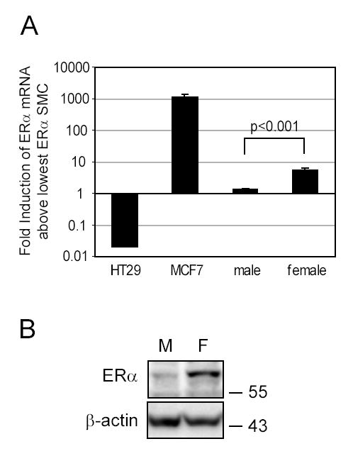
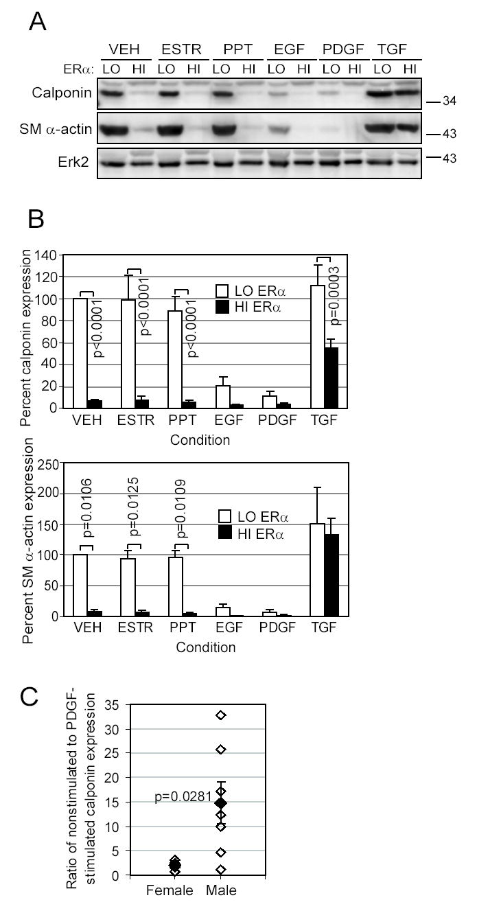
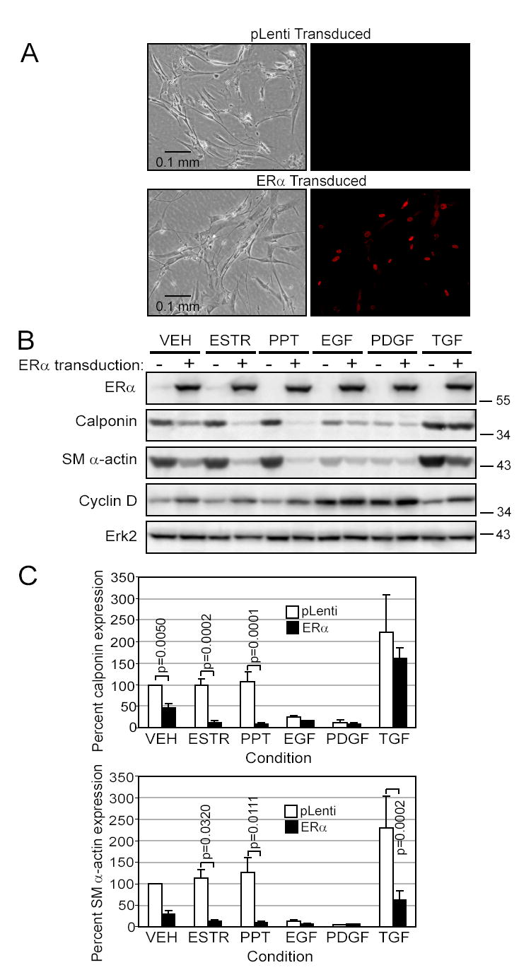
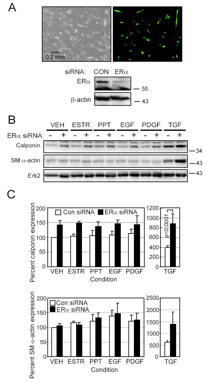
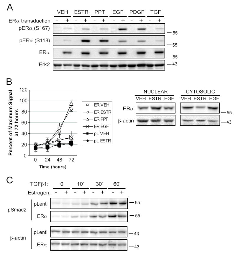
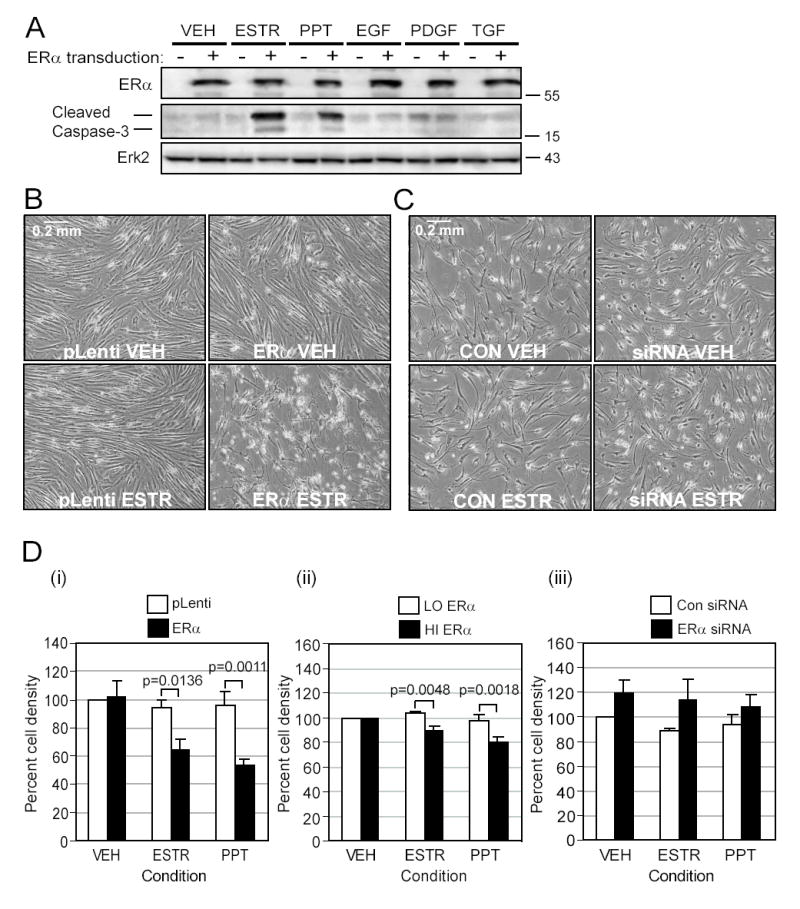
Women are at high risk of dying from unrecognized cardiovascular disease. Many differences in cardiovascular disease between men and women appear to be mediated by vascular smooth muscle cells (SMC). Because estrogen reduces the proliferation of SMC, we hypothesized that activation of estrogen receptor-alpha (ERalpha) by agonists or by growth factors altered SMC function. To determine the effect of growth factors, estrogen, and ERalpha expression on SMC differentiation, human aortic SMC were cultured in serum-free conditions for 10 days. SMC from men had lower spontaneous expression of ERalpha and higher levels of the differentiation markers calponin and smooth muscle alpha-actin than SMC from women. When SMC containing low expression of ERalpha were transduced with a lentivirus containing ERalpha, activation of the receptor by ligands or growth factors reduced differentiation markers. Conversely, inhibiting ERalpha expression by small interfering RNA (siRNA) in cells expressing high levels of ERalpha enhanced the expression of differentiation markers. ERalpha expression and activation reduced the phosphorylation of Smad2, a signaling molecule important in differentiation of SMC and initiated cell death through cleavage of caspase-3. We conclude that ERalpha activation switched SMC to a dedifferentiated phenotype and may contribute to plaque instability.


Conflict of interest statement

Conflict of Interest disclosure No conflict of interest exists among the authors and any company or advisory council relevant to this work.

Figures

Figure 1
Figure 1. Aortic SMC from female donors have more ERα than cells from male donors
A. Aortic SMC from five male and five female tissue donors were lysed after 5 days of starvation to analyze ERα mRNA levels by Real-Time PCR. ERα mRNA levels for SMC, the colon cancer cell line HT29 (negative control) and the ERα-positive breast cancer cell line MCF7 (positive control) were expressed as fold-induction above the cell population from the male SMC which consistently had the lowest levels of ERα expression. B. SMC expressing the lowest and highest levels of mRNA for ERα (M: male, F: female) were starved in basal media for 10 days and Western blotted for ERα expression. β-actin was detected as a loading control.
Figure 2
Figure 2. Differentiation of aortic SMC varies inversely to ERα expression
SMC with the highest (HI) or the lowest (LO) ERα expression were exposed for 10 days in EBM-PRF to VEH control, ESTR (10nmol/L), PPT (10nmol/L), EGF (10 ng/mL), PDGF-BB (10 ng/mL), or TGFβ1 (5 ng/mL). A. Western blotting was performed on protein lysates to determine expression of contractile proteins calponin and SM α-actin, and Erk2 as a loading control. A representative blot is shown. B. Protein expression was quantitated by densitometry, normalized by the Erk2 band, and stated as a percent of the low-ERα VEH control condition (□low-ERα SMC, ■high-ERα SMC, mean±SEM of 4 replicates). C. SMC from three male donors and three female donors were grown for 10 days in EBM-PRF with either PDGF-BB or VEH control. Western blotting was performed to detect calponin, and differentiation was calculated as a ratio of the intensity for the VEH-treated to the PDGF-treated samples (multiple replicates: ⋄n=6 female, n=7 male, ♦mean±SEM).
Figure 3
Figure 3. SMC dedifferentiate following transduction of ERα
SMC transduced with either an empty vector (pLenti) or ERα cDNA were exposed for 10 days in EBM-PRF with various stimuli as indicated in methods and Figure 2 A. Immunofluorescent staining of ERα illustrates the efficiency of transduction. B. Western blots demonstrate expression of ERα, loss of calponin and SM α-actin contractile proteins and upregulation of cyclin D1 in the ERα-transduced cells. C. Calponin and SM α-actin densitometry were expressed as a percent of the pLenti VEH control (□pLenti or ■ERα, mean±SEM of 4 (calponin) or 5 (SM α-actin)).
Figure 4
Figure 4. Reduction of ERα expression by siRNA induces differentiation
SMC expressing high levels of ERα were transfected with either empty vector or plasmid producing ERα siRNA. A. Transfection of pmaxGFP into high-ERα SMC illustrated the high transfection efficiency. Reduction in ERα protein levels by siRNA in cells starved for 10 days was demonstrated by Western blotting. β-actin was used as a loading control. B. Transfected cells were exposed for 10 days in EBM-PRF with stimuli as indicated in methods. Western blotting was performed to detect the differentiation markers calponin and SM α-actin. C. Calponin and SMα-actin densitometry as a percent of the empty vector VEH control condition was calculated (mean±SEM of 4 replicates, transfected with either □empty vector or ■ERα siRNA).
Figure 5
Figure 5. Ligand activation of ERα causes nuclear translocation and inhibition of Smad2 phosphorylation
SMC transduced with pLenti or ERα virus were stably selected with blasticidin and activated with the various stimuli as indicated previously. A. Western blots from cells activated for 20 minutes were sequentially immunoblotted with antibodies to phospho-ERα (S167), phospho-ERα (S118), ERα, and Erk2, with stripping of the membrane between antibodies. Shown is a representative figure from two experiments. B. Stably transduced cells were transfected with an estrogen response element reporter construct producing secreted alkaline phosphatase (SEAP) and were treated as described for up to three days. Aliquots of the media were analyzed for SEAP each day and luminescent signal was normalized as a percent of the maximum signal achieved on day 3 (mean±SEM of 4 experiments). Western blotting of nuclear and cytosolic lysates from SMC transduced with ERα and activated for 20 minutes as indicated demonstrated nuclear translocation of ERα when activated by ESTR. C. SMC stably transduced with pLenti or ERα were pre-incubated with ESTR (+) or ETOH (−) for 30 minutes, then activated with TGFβ1 for the time indicated. Phosphorylated Smad2 and β-actin as a loading control were detected by Western blotting (representative of two experiments).
Figure 6
Figure 6. ERα ligands cause apoptosis of aortic SMC
A. SMC virally transduced with either pLenti or pLenti-ERα were exposed for 10 days in EBM-PRF with various stimuli as indicated in methods. The cleaved or active form of caspase-3 was detected at 17 and 19kD by SDS-PAGE Western blots. B. Representative phase contrast photos were taken of SMC containing low ERα levels transduced with either pLenti or ERα and treated with VEH or ESTR for 10 days. C. Representative photos are shown of cells with high endogenous levels of ERα transfected with either control or ERα siRNA plasmid and treated with VEH or ESTR for 10 days. D. Phase contrast photos were quantitated for pixel density of live (gray) cells in the following groups: (i) pLenti- (□) or ERα-transduced cells (■) activated by ERα agonists (n=5); (ii) SMC natively expressing low ERα levels (□) or high ERα levels (■)(n=4); or (iii) SMC expressing high ERα levels transfected with empty plasmid (□) or siRNA plasmid (■) (n=4).

Comment in

References

    1. American Heart Association . Heart Disease and Stroke Statistics - 2005 Update. Dallas, Texas: 2005.
    1. Mendelsohn ME, Karas RH. Molecular and cellular basis of cardiovascular gender differences. Science. 2005;308:1583–1587. - PubMed
    1. Rossouw JE, Anderson GL, Prentice RL, LaCroix AZ, Kooperberg C, Stefanick ML, Jackson RD, Beresford SA, Howard BV, Johnson KC, Kotchen JM, Ockene J. Risks and benefits of estrogen plus progestin in healthy postmenopausal women: principal results From the Women’s Health Initiative randomized controlled trial. JAMA. 2002;288:321–333. - PubMed
    1. Hulley S, Grady D, Bush T, Furberg C, Herrington D, Riggs B, Vittinghoff E. Randomized trial of estrogen plus progestin for secondary prevention of coronary heart disease in postmenopausal women. Heart and Estrogen/progestin Replacement Study (HERS) Research Group. JAMA. 1998;280:605–613. - PubMed
    1. Grodstein F, Manson JE, Stampfer MJ. Hormone Therapy and Coronary Heart Disease: The Role of Time since Menopause and Age at Hormone Initiation. J Womens Health (Larchmt ) 2006;15:35–44. - PubMed

Publication types

MeSH terms