. 2005 Apr;1(1):53-8.
doi: 10.4161/auto.1.1.1589. Epub 2005 Apr 4.

Macrophages rapidly transfer pathogens from lipid raft vacuoles to autophagosomes

Affiliations

Macrophages rapidly transfer pathogens from lipid raft vacuoles to autophagosomes

Amal O Amer et al. Autophagy. 2005 Apr.

Abstract

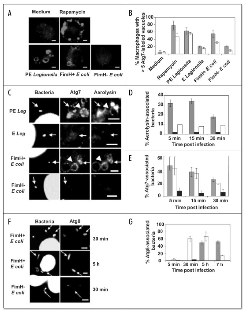
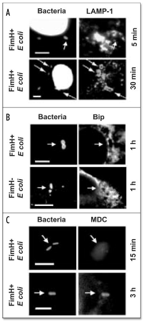
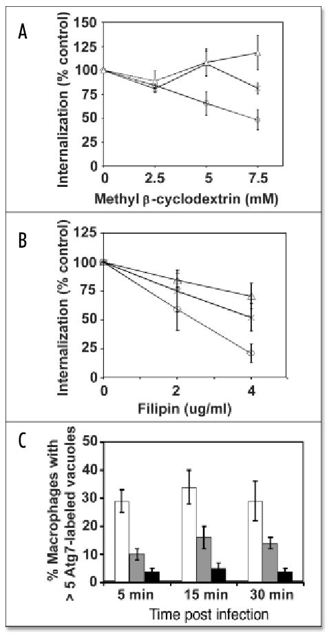
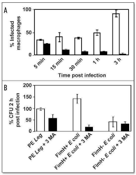
Macrophages activate autophagy as an immediate response to Legionella pneumophila infection, but what marks the pathogen phagosome as a target for the autophagy machinery is not known. Because a variety of bacteria, parasites, viruses, and toxins that associate with the endoplasmic reticulum enter host cells by a cholesterol-dependent route, we tested the hypothesis that autophagy is triggered when microbes engage components of lipid raft domains. As the intracellular respiratory pathogen L. pneumophila or the extracellular uropathogen FimH(+) Escherichia coli entered macrophages by a cholesterol-sensitive mechanism, they immediatezly resided in vacuoles rich in glycosylphosphatidylinositol moieties and the autophagy enzyme Atg7. As expected for autophagosomes, the vacuoles sequentially acquired the endoplasmic reticulum protein BiP, the autophagy markers Atg8 and monodansyl-cadaverine, and the lysosomal protein LAMP-1. A robust macrophage response to the pathogens was cholesterol-dependent, since fewer Atg7-rich vacuoles were observed when macrophages were pretreated with methyl-beta-cyclodextrin or filipin. A model in which macrophages exploit autophagy to capture pathogens within the lipid raft pathway for antigen presentation prior to disposal in lysosomes is discussed.


Figures

Figure 1
Figure 1
L. pneumophila and uropathogenic E. coli phagosomes colocalize with markers of the lipid raft and autophagy pathways. (A) Atg7 redistributed to vacuoles within 15 min of incubating macrophages with the autophagy stimulator rapamycin or with virulent PE L. pneumophila or FimH+ E. coli but not with medium or FimH E. coli. (B) The percent macrophages with > 5 Atg7-labeled vacuoles at 5 min (grey bars) and 15 min (white bars) after treatments described in (A); avirulent E L. pneumophila served as an additional negative control. Values are the mean percent of at least three independent experiments ± standard deviation. (C) PE L. pneumophila or FimH+ E. coli stimulated formation of vacuoles (arrowheads) and phagosomes (arrows) rich in Atg7 and GPI-molecules (aerolysin), but E L. pneumophila or FimH E. coli did not. (D) Phagosomes containing PE L. pneumophila (grey bars) retained Alexa 594-aerolysin, unlike those with E wild-type (black) or PE dotA mutants (white bars). Values are the mean percent of at least two independent experiments ± standard deviation. (E) Atg7 accumulated on phagosomes containing PE L. pneumophila (grey bars) or FimH+ E. coli (white bars), but not FimH E. coli (black bars). (F) FimH+ E. coli persisted in vacuoles that colocalized with Atg8, unlike FimH E. coli. Values are the mean percent of at least two independent experiments ± standard deviation. (G) Atg8 accumulated on phagosomes of PE L. pneumophila (grey bars) or FimH+ E. coli (white bars), but not FimH E. coli (black bars). Values are the mean percent of at least two independent experiments ± standard deviation. Scale bar, 10 μm.
Figure 2
Figure 2
Uropathogenic E. coli phagosomes acquire Bip, LAMP-1 and MDC. (A) DAPI-stained FimH+ E. coli vacuoles required 30 min to accumulate the late endosomal and lysosomal protein LAMP-1. (B) FimH+ but not FimH E. coli vacuoles colocalized with the ER protein Bip 1 h after infection. (C) The autophagosomal marker MDC accumulated in FimH+ E. coli vacuoles after 3 h of infection. E. coli were stained with NHS-carboxy-fluorescein in (B and C). Scale bar, 10 μm.
Figure 3
Figure 3
Perturbation of lipid rafts decreased pathogen entry and autophagosome formation. Methyl-β-cyclodextrin (A) or filipin (B) pretreatment for 1 h primarily decreased macrophage internalization of PE L. pneumophila (circles) but had moderate effect on uptake of dotA L. pneumophila (X) or DH5α E. coli (triangles). Percent internalization was quantified microscopically as: [(# intracellular bacteria 15 min post-infection)treated/(# intracellular bacteria 15 min post-infection)]untreated × 100. (C) Macrophages contained numerous Atg7-decorated vacuoles when infected by PE L. pneumophila (white bars), unless they were pretreated for 1 h with 5 mM methyl-β-cyclodextrin (grey) or 4 μg/ml filipin (black bars). Values are the mean percent of at least three independent experiments ± standard deviation.
Figure 4
Figure 4
Viability of pathogens in autophagosomes. (A) During a 3 h incubation period, FimH+ E. coli infected additional cells (white bars), whereas FimH E. coli were cleared (black bars), as judged microscopically by the percent of macrophages that contained intact bacteria. (B) Compared to control cultures (white bars), macrophages pretreated for 1 h with 3MA to inhibit autophagy (black bars) decreased the colony forming units (CFU) of pathogenic PE L. pneumophila and FimH+ E. coli, whereas FimH E. coli were rapidly killed by both treated and untreated macrophages. Percent CFU 2 h post-infection was calculated as [(intracellular CFU 2 h post-infection)/(intracellular CFU 5 min post-infection)] x 100. Values are the mean percent of at least two independent experiments ± standard deviation.

References

    1. Baorto DM, Gao Z, Malaviya R, Dustin ML, van der Merwe A, Lublin DM, Abraham SN. Survival of FimH-expressing enterobacteria in macrophages relies on glycolipid traffic. Nature. 1997;389:636–9. - PubMed
    1. Gatfield J, Pieters J. Essential role for cholesterol in entry of Mycobacteria into macrophages. Science. 2000;288:1647–50. - PubMed
    1. Manes S, del Real G, Martinez AC. Pathogens: Raft hijackers. Nat Rev Immunol. 2003;3:557–68. - PubMed
    1. Naroeni A, Porte F. Role of cholesterol and the ganglioside GM1 in entry and short term survival of Brucella suis in murine macrophages. Infect and Immun. 2002;70:1640–4. - PMC - PubMed
    1. Anderson HA, Chen Y, Norkin LC. Bound simian virus 40 translocates to caveolin-enriched membrane domains, and its entry is inhibited by drugs that selectively disrupt caveolae. Mol Biol Cell. 1996;7:1825–34. - PMC - PubMed

Publication types

MeSH terms

LinkOut - more resources