. 2006 Aug 9;25(15):3596-604.
doi: 10.1038/sj.emboj.7601248. Epub 2006 Jul 27.

Recruitment of TFIIH to the HIV LTR is a rate-limiting step in the emergence of HIV from latency

Affiliations

Recruitment of TFIIH to the HIV LTR is a rate-limiting step in the emergence of HIV from latency

Young Kyeung Kim et al. EMBO J. .

Abstract

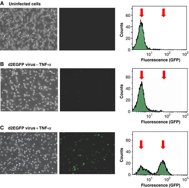
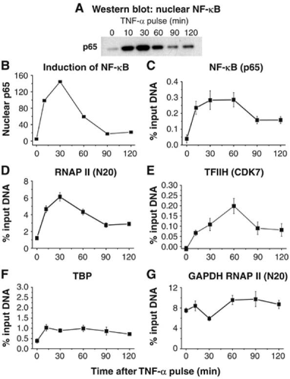
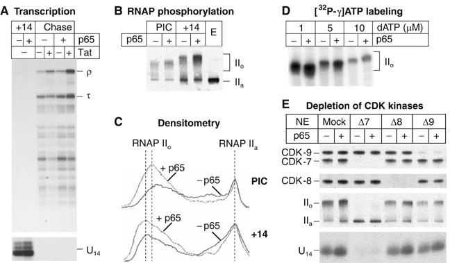
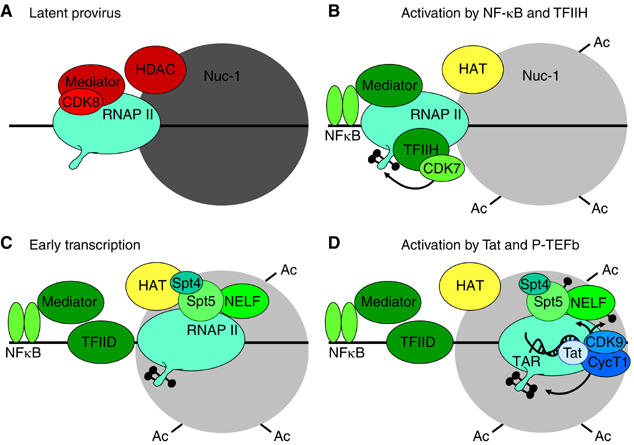
Latently infected cells rapidly initiate HIV transcription after exposure to signals that induce NF-kappaB. To investigate the role of TFIIH during HIV reactivation in vivo, we developed a population of Jurkat cells containing integrated, but transcriptionally silent, HIV proviruses. Surprisingly, the HIV promoter in unactivated Jurkat T cells is partially occupied and carries Mediator containing the CDK8 repressive module, TFIID and RNAP II that is hypophosphorylated and confined to the promoter region. Significantly, the promoter is devoid of TFIIH. Upon stimulation of the cells by TNF-alpha, NF-kappaB and TFIIH are rapidly recruited to the promoter together with additional Mediator and RNAP II, but CDK8 is lost. Detailed time courses show that the levels of TFIIH at the promoter fluctuate in parallel with NF-kappaB recruitment to the promoter. Similarly, recombinant p65 activates HIV transcription in vitro and stimulates phosphorylation of the RNAP II CTD by the CDK7 kinase module of TFIIH. We conclude that the recruitment and activation of TFIIH represents a rate-limiting step for the emergence of HIV from latency.


Figures

Figure 1
Figure 1
Induction of HIV gene expression in latently infected Jurkat T-cells by TNF-α. Populations of Jurkat T cells were infected with VSV-G pseudotyped vectors carrying the d2EGFP-reporter (short-lived GFP) and a wild-type Tat gene. The cells spontaneously shutdown transcription and enter latency. (A) Uninfected cells. (B) Latently infected Jurkat T cells prior to TNF-α treatment. (C) Latently infected Jurkat T-cells induced with TNF-α for 15 h. Left panels: Light micrographs of the cell population. Middle panels: Fluorescent micrographs. Right panels: Histogram of fluorescent cells obtained by FACS. Arrows indicate the mean positions of the negative and positive cell populations.
Figure 2
Figure 2
Distribution of RNA polymerase on proviral genomes before and after induction of NF-κB. (A) Map of lentiviral vector and locations of primers for ChIP analysis. The virus carries the d2EGFP gene (GFP) and a wild-type Tat gene. (B) ChIP assay using RNAP (N20) antibody to the RNAP large subunit showing distribution of RNA polymerase along proviral genome following TNF-α stimulation for 30 min, in the presence or absence of Tat. Data represent the average of at least four determinations±standard error of the mean.
Figure 3
Figure 3
The recruitment of TFIIH to HIV promoter is induced by NF-κB and leads to enhanced RNAP phosphorylation. Jurkat cells populations containing HIV proviruses carrying the C22G mutation in Tat (−Tat cells) and either a wild-type NF-κB binding site (WT) or a mutated NF-κB binding site (κB-Mut) were stimulated with TNF-α for 30 min and then analyzed by ChIP assays. The −116 to +4 region corresponding to the HIV promoter was amplified for this analysis. (A) Total RNAP II detected by the N20 antibody. (B) Phosphorylated RNAP II detected by the H14 antibody. (C) p65 (NF-κB). (D) TFIIH (CDK7; C-19). (E) TFIIH (p62). (F) TFIIH (p89). (G) TFIID (TBP). (H) Mediator recruitment detected using antibody to TRAP150. (I) CDK8 loss following NF-κB activation. Data represent the average of at least four determinations±standard error of the mean.
Figure 4
Figure 4
RNAP II is rapidly recruited to the HIV promoter following TNF-α induction of NF-κB. (A) Western blot of p65 in nuclear extracts at various times after exposure of the cells to TNF-α. (B) Densitometry plot of the gel. (C) ChIP assay showing fluctuating p65 levels at the HIV promoter. (D) RNAP II (N20 antibody). (E) CDK7 (FL-346 antibody). (F) TBP. (G) RNAP II at the GAPDH gene. Note that the recruitment of RNAP II and CDK7 occurs in parallel with p65 binding to the HIV LTR.
Figure 5
Figure 5
TFIIH is recruited to HIV promoter only in the presence of NF-κB. Jurkat cells containing HIV proviruses carrying either wild type (+Tat) or C22G mutant Tat (−Tat) genes were activated by treatment with TNF-α from between 10 and 120 min. (A) RNAP II at the promoter (−116 to +4) detected by the N20 antibody. (B) RNAP II downstream of TAR (+286 to +390) detected by the N20 antibody. (C) TFIIH (p89) levels at the promoter. (D) TFIIH (p89) levels downstream of TAR. (E) P-TEFb (CDK9) levels at the promoter. (F) P-TEF6 (CDK9) levels downstream of TAR. Note that TFIIH recruitment to the promoter fluctuates in parallel with RNAP II levels due to the entry and exit of NF-κB from the nucleus. Data represent the average of four determinations±standard error of the mean.
Figure 6
Figure 6
NF-κB p65 stimulates HIV transcription initiation and RNA polymerase II phosphorylation in vitro. (A) Transcription. PICs and complexes paused at +14 (+14) by performing elongation reactions in the absence of ATP were formed on immobilized templates in the absence (−) or presence (+) of 400 ng of p65. Chase reactions were performed in the absence (−) or presence (+) of 20 ng of Tat protein. RNA products were purified and analyzed by urea–PAGE followed by autoradiography. τ, transcription complexes paused at the terminator sequence; ρ, transcripts reaching the end of the template. (B) Immunoblot showing RNAP II in purified PICs and +14 complexes using the N20 antibody recognizing the N-terminal domain of the largest subunit of RNAP II. Nuclear extract (E) was used as a control to show the unphosphorylated polymerase (IIa). Extensive CTD phosphorylation induces a decrease in the mobility of the protein on SDS–PAGE (IIo). (C) Quantitative analysis of the immunoblots showed in (B) by densitometry. (D) 32P-labeling of PICs. PICs were assembled in the absence (−) or presence (+) of p65, washed and incubated with 10 μCi of [γ-32P]ATP and an increasing concentration of unlabeled dATP, as indicated. The purified radioactive complexes were transferred onto a nitrocellulose membrane, and analyzed by autoradiography. (E) Immunodepletion of CDK8 or CDK9 does not prevent p65-induced stimulation of CTD phosphorylation. Nuclear extracts from Jurkat cells were immunodepleted with control rabbit immunoglobulins (Mock), anti-CDK7 (Δ7) anti-CDK8 (Δ8) or anti-p-TEFb (Δ9) antibodies. (Top panels) Western-blot of the transcription complexes paused at +14. Depletion of CDK8 or p-TEFb did not affect significantly the level of RNAP II CTD phosphorylation, whereas CDK7 depletion completely abolished both the basal and the p65-activated phosphorylation of RNAP II. IIa, unphosphorylated Pol II; IIo, hyperphosphorylated Pol II. (Bottom panel) Transcription reactions in depleted extracts. RNA products were purified from the transcription complexes paused at +14 and analyzed by urea–PAGE.
Figure 7
Figure 7
Model for activation of transcription from latent proviruses by NF-κB. (A) Latent provirus. PICs contain Mediator that has been inactivated by the CDK8 repressive module, and lack TFIIH. Initiation is further restricted by the nonacetylated nucleosome 1 (Nuc-1). (B) Activation by NF-κB and TFIIH. Following cellular activation, NF-κB enters the nucleus and binds to the HIV LTR. NF-κB causes the CDK8 module to dissociate from the Mediator and leads to the recruitment of TFIIH, which is then able to phosphorylate the RNAP II CTD. Histone acetyltransferases (HAT) are recruited and acetylate Nuc-1. (C) Early transcription. Elongation is restricted by DSIF (a complex Spt5 and Spt4) and NELF. These negative factors do not impose an absolute block to elongation but lead to the early dissociation of RNAP II from the template in the absence of Tat. (D) Activation by Tat and P-TEFb. After the transcription through the TAR element, Tat and P-TEFb (the CDK9 and CycT1 complex) are recruited to the elongation complex. This activates the CDK9 kinase and leads to phosphorylation of the CTD of RNA polymerase II, Spt5 and the RD subunit of NELF. The phosphorylation of NELF leads to its release and enhanced RNAP processivity. The presence of hyperphosphorylated RNAP II and Spt5 allows transcription of the full HIV genome and read through a wide variety of blocks to transcription elongation. Several important transcription factors known to be present at the HIV LTR including SP1 and the TAFs have been omitted for clarity.

Similar articles

Cited by

References

    1. Alcami J, de Lera TL, Folgueira L, Pedraza M-A, Jacqué J-M, Bachelerie F, Noriega AR, Hay RT, Harrich D, Gaynor RB, Virelizier J-L, Arenzana-Seisdedos F (1995) Absolute dependence on kB responsive elements for initiation and Tat-mediated amplification of HIV transcription in blood CD4T lymphocytes. EMBO J 14: 1552–1560 - PMC - PubMed
    1. Barboric M, Nissen RM, Kanazawa S, Jabrane-Ferrat N, Peterlin BM (2001) NF-kB binds P-TEFb to stimulate transcriptional elongation by RNA polymerase II. Mol Cell 8: 327–337 - PubMed
    1. Barboric M, Peterlin BM (2005) A new paradigm in eukaryotic biology: HIV Tat and the control of transcriptional elongation. PLoS Biol 3: e76. - PMC - PubMed
    1. Bieniasz PD, Grdina TA, Bogerd HP, Cullen BR (1998) Recruitment of a protein complex containing Tat and cyclin T1 to TAR governs the species specificity of HIV-1 Tat. EMBO J 17: 7056–7065 - PMC - PubMed
    1. Blau J, Xiao H, McCracken S, O'Hare P, Greenblatt J, Bentley D (1996) Three functional classes of transcriptional activation domains. Mol Cell Biol 16: 2044–2055 - PMC - PubMed

Publication types