. 2006 Aug 9;25(15):3546-55.
doi: 10.1038/sj.emboj.7601239. Epub 2006 Jul 27.

Rapamycin activates Tap42-associated phosphatases by abrogating their association with Tor complex 1

Affiliations

Rapamycin activates Tap42-associated phosphatases by abrogating their association with Tor complex 1

Gonghong Yan et al. EMBO J. .

Abstract

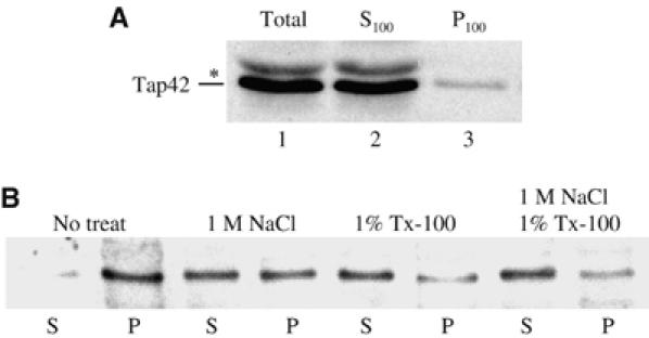
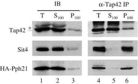
In Saccharomyces cerevisiae, the Tap42-phosphatase complexes are major targets of the Tor kinases in the rapamycin-sensitive signaling pathway. The immunosuppressive agent, rapamycin, induces a prompt activation of the Tap42-associated phosphatases, which is vitally important in Tor-mediated transcriptional regulation. However, the mechanism for the rapid phosphatase activation is poorly understood. In this study, we show that the Tap42-phosphatase complexes exist mainly on membrane structures through their association with Tor complex 1 (TORC1). Rapamycin abrogates this association and releases the Tap42-phosphatase complexes into the cytosol. Disassembly of the Tap42-phosphatase complexes occurs subsequently, following the release but at a much slower rate, presumably caused by Tap42 dephosphorylation. Release of the Tap42-phosphatase complexes from membrane structures also occurs when cells are deprived of nutrient. These findings suggest that the association of the Tap42-phosphatase complexes with TORC1 represents an important mechanism by which nutrient controls Tor signaling activity. In addition, our data support a model in which rapamycin acts not by inhibiting the kinase activity of Tor but by disrupting its interaction with downstream targets.


Figures

Figure 1
Figure 1
Tap42 associates with membrane structures. Extracts (Total) from exponentially growing cells (Y959) were partitioned into soluble (S100) and membrane (P100) fractions. (A) The presence of Tap42 in each fraction was analyzed by immunoblotting. (B) The membrane fraction was resuspended and treated with the indicated reagents for 30 min on ice followed by centrifugation at 100 000 g. The levels of Tap42 in the supernatant (S) and pellet (P) were analyzed by immunoblotting. ‘*' in this and other figures denotes a nonspecific protein.
Figure 2
Figure 2
Tap42 is in complex with TORC1. (A) Extracts from cells expressing HA-Tor2 (BY2470) were fractionated into soluble (S100) and membrane (P100) fractions. The distribution of Tap42 and HA-Tor2 in the fractions was determined by immunoblotting (IB). Both fractions were treated with Triton X-100, and precipitated with (α-Tap42) or without (mock) anti-Tap42 antibody. The presence of Tap42 and HA-Tor2 in the precipitates was analyzed by immunoblotting (IP). (B) Extracts from cells expressing HA-Tor1 (Y990) were fractionated into soluble (S100) and membrane (P100) fractions, and the distribution of Tap42 and HA-Tor1 in the fractions was analyzed by immunoblotting (IB). Both fractions were treated with Triton X-100 and precipitated with (α-Tap42) or without (mock) anti-Tap42 antibody. The presence of Tap42 and HA-Tor1 in the precipitates was determined by immunoblotting (IP). (C) The membrane fraction (P100) of the cells expressing HA-Kog1 (Y1032) was solubilized with Triton X-100, and precipitated with (α-Tap42) or without (mock) anti-Tap42 antibody. The presence of Tap42 and HA-Kog1 in the precipitate was analyzed by immunoblotting (IP). (D) The membrane fraction (P100) of cells expressing HA-Avo1 (Y1031) was solubilized with Triton X-100 and precipitated with (α-Tap42) or without (mock) anti-Tap42 antibody. The presence of Tap42 and HA-Avo1 in the precipitate was analyzed by immunoblotting (IP). In panels C and D, the amount of P100 in lane 1 represents 10% of the total input for the precipitation shown in lanes 2 and 3.
Figure 3
Figure 3
The association of Tap42 with Tor2 is sensitive to rapamycin. Exponentially growing cells expressing HA-TOR2 (BY2470) were treated with (+) or without (−) 200 nM of rapamycin for 1 h and lysed. (A) Cell extracts were partitioned into soluble and membrane fractions. The distribution of Tap42 (upper panel) and HA-Tor2 (lower panel) in the total extracts (T) and membrane fraction (P100) was determined by immunoblotting. (B) Extracts from cells (BY2470) treated with (+) or without (−) rapamycin were treated with Triton X-100, and precipitated with anti-Tap42 antibody. The presence of Tap42 and HA-Tor2 in the precipitates was determined by immunoblotting.
Figure 4
Figure 4
Tap42 is phosphorylated only when it is associated with TORC1. (A) Exponentially growing cells (Y959) were metabolically labeled with 32P and lysed. Cell extracts were partitioned into soluble and membrane fractions. The total cell extracts (T), soluble (S100) and the resuspended membrane (P100) fractions were boiled in the presence of SDS, diluted with buffer containing Triton X-100 and then precipitated with anti-Tap42 antibody. (B) Exponentially growing cells (Y959) were metabolically labeled with 32P followed by treatment with rapamycin. An aliquot of cells was removed at the indicated time point after addition of the drug and processed as described above. The precipitated Tap42 protein was detected by immunoblotting (IB) and the incorporation of the radiolabel in the protein was visualized by autoradiography (32P).
Figure 5
Figure 5
The Tap42–phosphatase complexes are associated with TORC1. (A) Cells expressing HA-PPH21 (Y162) were grown to early log phase and treated with (+) or without (−) 200 nM of rapamycin for 1 h. Cells were lysed and extracts were centrifuged at 100 000 g to obtain membrane fractions. The distribution of HA-Pph21 (upper panel), Sit4 (middle panel) and Tap42 (lower panel) in the total cell extracts (Total) and membrane fractions (P100) in response to rapamycin treatment was determined by immunoblotting. (B) Wild-type cells (Y959) and cells expressing myc-Pph21 together with HA-TOR2 (Y992) were grown to early log phase and lysed. The membrane fraction of the cell extracts was solubilized with Triton X-100 and precipitated with anti-HA antibody. The presence of HA-Tor2, Tap42, myc-Pph21 and Sit4 in the precipitates was determined by immunoblotting.
Figure 6
Figure 6
The association of the Tap42–phosphatase complexes with membrane is resistant to rapamycin in tap42-11 cells. Wild-type (Y959) and tap42-11 cells (Y176) were grown to early log phase and treated with (+) or without (−) 200 nM of rapamycin for 1 h. Cells were lysed and extracts were centrifuged at 100 000 g to obtain membrane fractions. The presence of Tap42 in the total cell extracts (Total) and that of Tap42 and Sit4 in the membrane fractions (P100) in response to rapamycin treatment was determined by immunoblotting.
Figure 7
Figure 7
The Tap42–phosphatase complexes exist mainly on membrane. Cells expressing HA-Pph21 (Y162) were grown to early log phase and lysed. Cell lysates were partitioned into soluble and membrane fractions by centrifugation at 100 000 g. The presence of Tap42, Sit4 and HA-Pph21 in the fractions was determined by immunoblotting (IB). The total extracts (T), soluble (S100) and membrane (P100) fractions were treated with Triton X-100, and precipitated with anti-Tap42 antibody. The presence of Tap42, Sit4 and HA-Pph21 in the precipitates from each fraction was determined by immunoblotting (IP). The amount of Tap42 shown in the IB panel represents half of the input used for the precipitation shown in the IP panel. The amount of Sit4 and HA-Pph21 shown in the IB panel represents 10% of the input used for the precipitation shown in the IP panel.
Figure 8
Figure 8
Rapamycin induces rapid redistribution of the Tap42–phosphatase complexes into the cytosol. Cells of strain Y162 were grown to early log phase and treated with 200 nM of rapamycin. Aliquots of cells were removed from the culture at the indicated time points after addition of the drug, and lysed immediately. The cells lysates were partitioned into membrane and soluble fractions by centrifugation at 100 000 g. (A) The amount of Tap42, Sit4 and HA-Pph21 associated with the membrane fraction from samples taken at the indicated time points after addition of rapamycin was determined by immunoblotting. (B) The soluble fractions from samples taken at the indicated time points were immunoprecipitated with anti-Tap42 antibody, and the amount of Tap42, Sit4 and HA-Pph21 in the precipitates was determined by immunoblotting. (C) The level of Tap42, Sit4 and HA-Pph21 in the total cell extracts at the indicated time points after rapamycin treatment was determined by immunoblotting. The first lane, depicted as ‘U', shows the result of the untreated cells.
Figure 9
Figure 9
Nutrient starvation causes a rapid release of the Tap42–phosphatase complexes from TORC1. Exponentially growing yeast cells (Y992) were shifted from YP medium to water. Aliquots of cells were collected at the indicated time points after the shift and lysed. The levels of Tap42, HA-Tor2, Sit4 and myc-Pph21 in the cell extracts were determined with immunoblotting (extract). Cell extracts were precipitated with Tap42 antibody. The amount of Tap42, HA-Tor2, Sit4 and myc-Pph21 in the precipitates was determined by immunoblotting (IP).
Figure 10
Figure 10
Model for rapamycin-induced phosphatase activation. The Tap42–phosphatase complexes exist mainly on membrane structures through association with TORC1. Rapamycin abrogates the association and releases the complexes into the cytosol, where they become active. Disassembly of the Tap42 complexes occurs when Tap42 is dephosphorylated by the PP2A holoenzyme in the cytosol, which terminates the Tap42-dependent phosphatase activity. Pase: phosphatase.

References

    1. Barbet NC, Schneider U, Helliwell SB, Stansfield I, Tuite MF, Hall MN (1996) TOR controls translation initiation and early G1 progression in yeast. Mol Biol Cell 7: 25–42 - PMC - PubMed
    1. Beck T, Hall MN (1999) The TOR signalling pathway controls nuclear localization of nutrient-regulated transcription factors. Nature 402: 689–692 - PubMed
    1. Cardenas ME, Cutler NS, Lorenz MC, Di Como CJ, Heitman J (1999) The TOR signaling cascade regulates gene expression in response to nutrients. Genes Dev 13: 3271–3279 - PMC - PubMed
    1. Di Como CJ, Arndt KT (1996) Nutrients, via the Tor proteins, stimulate the association of Tap42 with type 2A phosphatases. Genes Dev 10: 1904–1916 - PubMed
    1. Duvel K, Santhanam A, Garrett S, Schneper L, Broach JR (2003) Multiple roles of Tap42 in mediating rapamycin-induced transcriptional changes in yeast. Mol Cell 11: 1467–1478 - PubMed

Publication types

MeSH terms

Substances