

Preventive effects of chitosan on peritoneal adhesion in rats
- PMID: 16874876
- PMCID: PMC4125651
- DOI: 10.3748/wjg.v12.i28.4572
Preventive effects of chitosan on peritoneal adhesion in rats
Abstract
Aim: To study the effects of chitosan gel and blending chiston/gelatin film on preventing peritoneal adhesion in rats.
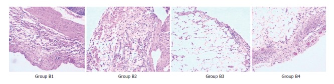
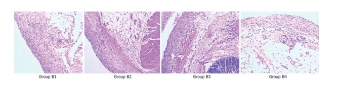
Methods: SD rats were randomly divided into 2 groups, group A treated with chitosan gel and group B with blending chiston/gelatin film. In group A, rats were randomly subdivided into 3 subgroups as groups A1, A2 and A3, and different methods were used to induce peritoneal adhesions at the dead end of vermiform process in each group as follows: Group A1 with trauma, A2 with talc powder and A3 with ligation of blood vessel. In each subgroup, rats were redivided into control group and experimental group whose treated vermiform processes were respectively coated with chitosan gel and normal saline immediately after the adhesion-induced treatments. In group B, all the rats received traumatic adhesion-induced treatments and then were randomly divided into 4 groups (groups B1, B2, B3, B4). Group B1 served as control group and were coated with normal saline in the vermiform processes immediately after the treatments, and groups B2, B3 and B4 with 100% chitosan film, chitosan film containing 10% gelatin and chiston film containing 50% gelatin, respectively. At 2 and 4 wk after the above treatments, half of the rats in each terminal group were belly opened, and the peritoneal adhesive situation was graded and histopathological changes were examined.
Results: (1) In group A, regarding peritoneal adhesion situation: At both 2 and 4 wk after the treatments, for groups A1 and A3, the adhesive grades of experimental groups were significantly lower than those of the control group (2 wk: H = 4.305, P < 0.05 for A1, H = 6.743, P < 0.01 for A3; 4 wk: H = 4.459, P < 0.05 for A1, H = 4.493, P < 0.05 for A3). However, of group A2, there was no significant difference between the experimental and control groups (2 wk: H = 0.147, P > 0.05; 4 wk: H = 1.240, P > 0.05). Regarding pathological changes: In groups A1 and A3, the main pathological change was fibroplasia. In group A2, the main changes were massive foreign-body giant cell reaction and granuloma formation with fibroplasia of different degrees. (2) In group B, regarding degradation of film: With increase of the blended gelatin concentration, degrading speed of the film accelerated significantly. Regarding peritoneal adhesion situation: At both 2 and 4 wk after the treatments, the adhesive grades of B1 were the lowest among the four subgroups of B (2 wk: H = 29.679, P < 0.05; 4 wk: H = 18.791, P < 0.05). At 2 wk after the treatments, the grades of group B2 were significantly lower than that of groups B3 and B4 (H = 4.025, P < 0.05 for B2 vs B3; H = 4.361, P < 0.05 for B2 vs B4). At 4 wk, there were no significant differences of the grades between groups B2, B3 and B4. Regarding pathological changes: Inflammatory cell infiltration and fibroplastic proliferation were observed in the local treated serous membranes, which was the mildest in group B1. Slight foreign-body giant cell reactions were also found in groups B2, B3, and B4.
Conclusion: (1) Chitosan gel has preventive effect on traumatic or ischemic peritoneal adhesion, but no obvious effect on foreign body-induced peritoneal adhesion. (2) Chitosan film may exacerbate the peritoneal adhesion. Blending with gelatin to chitosan film can accelerate the degradation of the film, but can simultaneously facilitate the formation of peritoneal adhesion.
Figures
References
-
- Hirano S. Chitin biotechnology applications. Biotechnol Annu Rev. 1996;2:237–258. - PubMed
-
- Shigemasa Y, Minami S. Applications of chitin and chitosan for biomaterials. Biotechnol Genet Eng Rev. 1996;13:383–420. - PubMed
-
- Kennedy R, Costain DJ, McAlister VC, Lee TD. Prevention of experimental postoperative peritoneal adhesions by N,O-carboxymethyl chitosan. Surgery. 1996;120:866–870. - PubMed
-
- Phillips RK, Dudley HA. The effect of tetracycline lavage and trauma on visceral and parietal peritoneal ultrastructure and adhesion formation. Br J Surg. 1984;71:537–539. - PubMed
-
- Holmdahl L, Eriksson E, al-Jabreen M, Risberg B. Fibrinolysis in human peritoneum during operation. Surgery. 1996;119:701–705. - PubMed
MeSH terms
Substances
LinkOut - more resources
Full Text Sources
Other Literature Sources
Medical
