

Coexistence of esophageal superficial carcinoma and multiple leiomyomas: A case report
- PMID: 16874880
- PMCID: PMC4125655
- DOI: 10.3748/wjg.v12.i28.4588
Coexistence of esophageal superficial carcinoma and multiple leiomyomas: A case report
Abstract
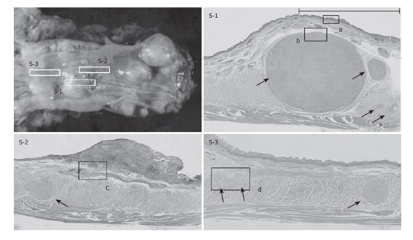
Leiomyomas are the most common benign tumors of the esophagus. They usually occur as a single lesion or as two or three nodules. Only two cases of esophageal multiple leiomyomas comprising more than 10 nodules have been reported previously. Moreover, there have been few reports of esophageal squamous cell carcinoma overlying submucosal tumors. We describe a 71-year-old man who was diagnosed as having a superficial esophageal cancer coexisting with two or three leiomyoma nodules. During surgery, 10 or more nodules that had not been evident preoperatively were palpable in the submucosal and muscular layers throughout the esophagus. As intramural metastasis of the esophageal cancer was suspected, we considered additional lymphadenectomy, but had to rule out this option because of the patient's severe anoxemia. Microscopic examination revealed that all the nodules were leiomyomas (20 lesions, up to 3 cm in diameter), and that invasion of the carcinoma cells was limited to the submucosal layer overlying a relatively large leiomyoma. This is the first report of superficial esophageal cancer coexisting with numerous solitary leiomyomas. Multiple minute leiomyomas are often misdiagnosed as intramural metastasis, and a leiomyoma at the base of a carcinoma lesion can also be misdiagnosed as tumor invasion. The present case shows that accurate diagnosis is required for the management of patients with coexisting superficial esophageal cancer and multiple leiomyomas.
Figures


References
-
- Kramer MD, Gibb SP, Ellis FH Jr. Giant leiomyoma of esophagus. J Surg Oncol. 1986;33:166–169. - PubMed
-
- Postlethwait RW, Musser AW. Changes in the esophagus in 1,000 autopsy specimens. J Thorac Cardiovasc Surg. 1974;68:953–956. - PubMed
-
- Takubo K, Nakagawa H, Tsuchiya S, Mitomo Y, Sasajima K, Shirota A. Seedling leiomyoma of the esophagus and esophagogastric junction zone. Hum Pathol. 1981;12:1006–1010. - PubMed
-
- Seremetis MG, Lyons WS, deGuzman VC, Peabody JW Jr. Leiomyomata of the esophagus. An analysis of 838 cases. Cancer. 1976;38:2166–2177. - PubMed
-
- Bradford ML, Mahon HW, Grow JB. Mediastinal cysts and tumors. Surg Gynecol Obstet. 1947;85:467–491. - PubMed
Publication types
MeSH terms
LinkOut - more resources
Full Text Sources
Medical
Research Materials
