. 2006 Sep;4(9):e268.
doi: 10.1371/journal.pbio.0040268.

c-Type cytochrome-dependent formation of U(IV) nanoparticles by Shewanella oneidensis

Affiliations

c-Type cytochrome-dependent formation of U(IV) nanoparticles by Shewanella oneidensis

Matthew J Marshall et al. PLoS Biol. 2006 Sep.

Abstract

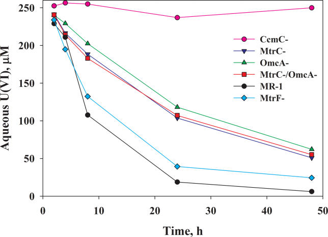
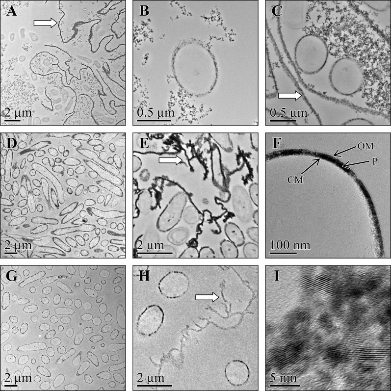
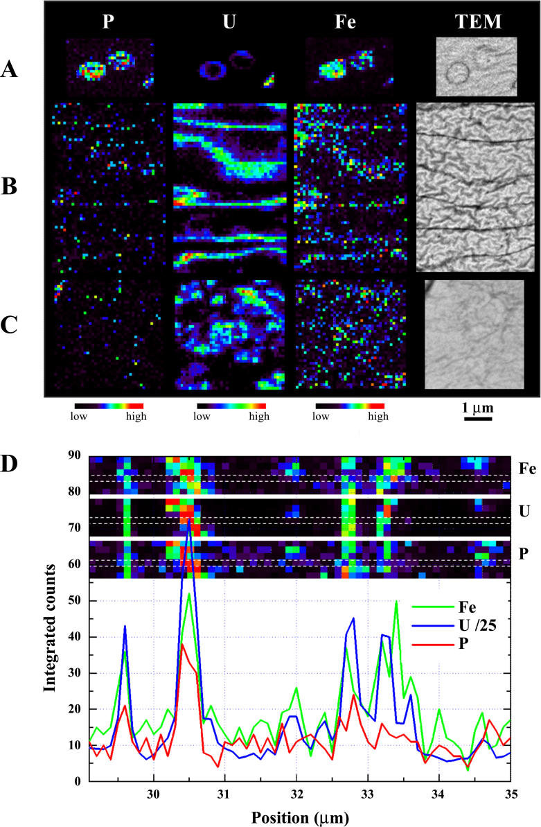
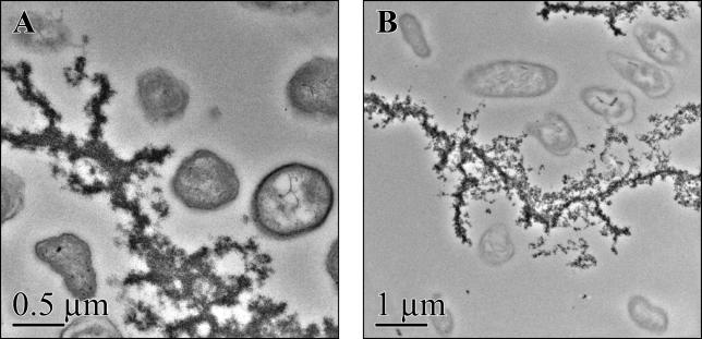
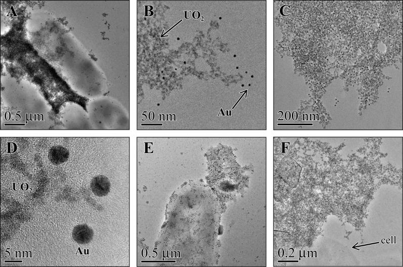
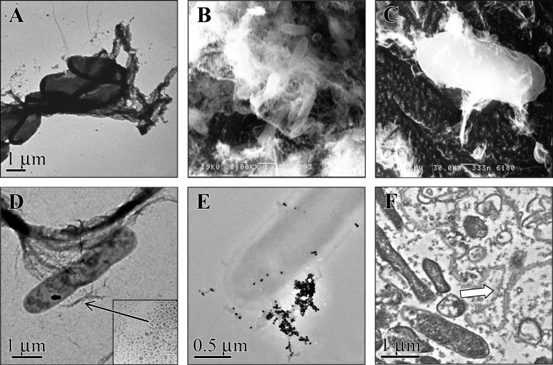
Modern approaches for bioremediation of radionuclide contaminated environments are based on the ability of microorganisms to effectively catalyze changes in the oxidation states of metals that in turn influence their solubility. Although microbial metal reduction has been identified as an effective means for immobilizing highly-soluble uranium(VI) complexes in situ, the biomolecular mechanisms of U(VI) reduction are not well understood. Here, we show that c-type cytochromes of a dissimilatory metal-reducing bacterium, Shewanella oneidensis MR-1, are essential for the reduction of U(VI) and formation of extracellular UO(2) nanoparticles. In particular, the outer membrane (OM) decaheme cytochrome MtrC (metal reduction), previously implicated in Mn(IV) and Fe(III) reduction, directly transferred electrons to U(VI). Additionally, deletions of mtrC and/or omcA significantly affected the in vivo U(VI) reduction rate relative to wild-type MR-1. Similar to the wild-type, the mutants accumulated UO(2) nanoparticles extracellularly to high densities in association with an extracellular polymeric substance (EPS). In wild-type cells, this UO(2)-EPS matrix exhibited glycocalyx-like properties and contained multiple elements of the OM, polysaccharide, and heme-containing proteins. Using a novel combination of methods including synchrotron-based X-ray fluorescence microscopy and high-resolution immune-electron microscopy, we demonstrate a close association of the extracellular UO(2) nanoparticles with MtrC and OmcA (outer membrane cytochrome). This is the first study to our knowledge to directly localize the OM-associated cytochromes with EPS, which contains biogenic UO(2) nanoparticles. In the environment, such association of UO(2) nanoparticles with biopolymers may exert a strong influence on subsequent behavior including susceptibility to oxidation by O(2) or transport in soils and sediments.


Figures

Figure 1
Figure 1. U(VI) Reduction Kinetics by S. oneidensis MR-1 and Cytochrome Mutant Cells
The reduction of 250 μM U(VI) was determined for MR-1, a mutant lacking all c-type cytochromes (CcmC ), single cytochrome deletion mutants (MtrC , OmcA , or MtrF ), and a double cytochrome deletion mutant (MtrC /OmcA ). Lines represent the mean data from representative experiments.
Figure 2
Figure 2. UO 2 Localization in S. oneidensis MR-1 Wild-type and Cytochrome-Deficient Mutants
TEM images prepared from cell suspensions incubated with 250 μM uranyl acetate and 10 mM lactate for 24 h. The localization of UO 2 by MR-1 (A, B) was compared to OmcA (C), MtrC (D–F), and MtrC /OmcA (G, H). High-resolution image of extracellular UO 2 nanoparticles showing d-lines values consistent to previous patterns of biogenic and synthetic UO 2 (I). The UO 2-EPS is designated by the arrows. Locations of the cell membrane (CM), periplasm (P), and outer membrane (OM) are shown.
Figure 3
Figure 3. Synchrotron XRF Microscopy of the Elemental Concentration Gradients in Association with S. oneidensis MR-1 Cells
False-color micro-XRF maps of qualitative spatial distributions and concentration gradients of P, U, and Fe in and around MR-1 cells. Cells are shown after incubation with 250 μM U(VI) for 24 h in standard assay conditions (A). The extracellular UO 2 precipitates associated with EPS (B) and diffuse extracellular UO 2 nanoparticles (C) observed in MR-1 samples were also evaluated for elemental composition. The scanned regions are represented with each corresponding thin-section TEM image. (D) The UO 2-EPS features seen in (B) were scanned vertically six times longer per point, and the pixel intensity (identified between the dashed lines) was plotted for each element. Although this image is of a smaller area and has a smaller number of features in its field of view, the increased measurement time provides more robust statistics and further supports co-localization of the elements.
Figure 4
Figure 4. Heme Staining of Extracellular Cytochromes from S. oneidensis MR-1
TEM images of thin sections of MR-1 incubated for 24 h with 100 μM U(VI) and stained for the presence of heme. Samples were incubated with DAB and developed with H 2O 2 (A) or with DAB but not developed with H 2O 2 (B) prior to embedding. Heme-containing proteins detected in (A) were shown in close association with the undeveloped UO 2-EPS seen in (B).
Figure 5
Figure 5. Immune-Localization of MtrC, OmcA, and MtrB with Extracellular UO 2 from S. oneidensis MR-1
Whole-mount TEM images of MR-1 incubated with 100 μM U(VI) for 24 h and reacted with specific antibodies to MtrC (A, B), OmcA (C, D), or MtrB (E). High-resolution image of nanocrystalline UO 2 associated with the extracellular matrix and the 5-nm particles of colloidal gold (Au) (B, D). Extracellular matrix and cell labeled with colloidal gold in the absence of specific antibody (F).
Figure 6
Figure 6. Extracellular Structure of S. oneidensis MR-1
Whole mounts of MR-1 incubated with 250 μM U(VI) for 24 h prior to visualization by TEM (A) or incubated in defined media and visualized by cryo-HRSEM (B, C). Whole-mount TEM of cells incubated with 1 mM fumarate added as the electron acceptor in place of U(VI) and reacted with positive charged colloidal nanogold particles (D) to help determine surface charge of the EPS matrix or the glycoconjugate-specific lectin, ConA, complexed with 40-nm particles of colloidal gold (E). High-resolution image of 1.4-nm gold nanoparticles (D inset). Thin-section TEM images of MR-1 incubated for 24 h with 1 mM fumarate prior to ruthenium red staining to visualize extracellular EPS (F). The ruthenium red-EPS is designated by the arrow.

Comment in

References

    1. Lovley DR, Holmes DE, Nevin KP. Dissimilatory Fe(III) and Mn(IV) reduction. Adv Microb Physiol. 2004;49:219–286. - PubMed
    1. Nealson KH, Saffarini D. Iron and manganese in anaerobic respiration: Environmental significance, physiology, and regulation. Annu Rev Microbiol. 1994;48:311–343. - PubMed
    1. Rusin PA, Quintana L, Brainard JR, Strietelmeier BA, Tait CD, et al. Solubilization of plutonium hydrous oxide by iron-reducing bacteria. Environ Sci Technol. 1994;28:1686–1690. - PubMed
    1. Carpentier W, Sandra K, De Smet I, Brige A, De Smet L, et al. Microbial reduction and precipitation of vanadium by Shewanella oneidensis . Appl Environ Microbiol. 2003;69:3636–3639. - PMC - PubMed
    1. Liu C, Gorby YA, Zachara JM, Fredrickson JK, Brown CF. Reduction kinetics of Fe(III), Co(III), U(VI), Cr(VI), and Tc(VII) in cultures of dissimilatory metal-reducing bacteria. Biotechnol Bioeng. 2002;80:637–649. - PubMed

Publication types

MeSH terms