

Transverse propagation in an expanded PSpice model for cardiac muscle with gap-junction ion channels
- PMID: 16875501
- PMCID: PMC1559629
- DOI: 10.1186/1475-925X-5-46
Transverse propagation in an expanded PSpice model for cardiac muscle with gap-junction ion channels
Abstract
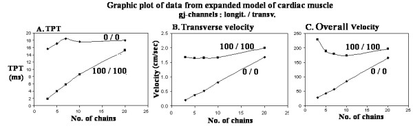
Transverse propagation was previously found to occur in a two-dimensional model of cardiac muscle using the PSpice software program for electronic circuit design and analysis. Longitudinal propagation within each chain, and transverse propagation between parallel chains, occurred even when there were no gap-junction (g-j) channels inserted between the simulated myocardial cells either longitudinally or transversely. In those studies, there were pronounced edge (boundary) effects and end-effects even within single chains. Transverse velocity increased with increase in model size. The present study was performed to examine boundary effects on transverse propagation velocity when the length of the chains was held constant at 10 cells and the number of parallel chains was varied from 3 to 5, to 7, to 10, and to 20. The number of g-j channels was either zero, both longitudinally and transversely (0/0), or 100/100. Some experiments were also made at 100/0, 1/1, and 10/10. Transverse velocity and overall velocity (both longitudinal and transverse components) was calculated from the measured total propagation time (TPT), i.e., the elapsed time between when the first action potential (AP) and the last AP crossed the zero potential level. The transverse g-j channels were placed only at the ends of each chain, such that propagation would occur in a zigzag pattern. Electrical stimulation was applied intracellularly between cells A1 and A2. It was found that, with no g-j channels (0/0), overall velocity increased almost linearly when more and more chains were placed in parallel. In contrast, with many g-j channels (100/100), there was a much flatter relationship between overall velocity and number of parallel chains. The difference in velocities with 0/0 channels and 100/100 channels was reduced as the number of chains was increased. In conclusion, edges have important effects on propagation velocity (overall and transverse) in cardiac muscle simulations.
Figures


Similar articles
-
Gap-junction channels inhibit transverse propagation in cardiac muscle.Biomed Eng Online. 2005 Jan 28;4:7. doi: 10.1186/1475-925X-4-7. Biomed Eng Online. 2005. PMID: 15679888 Free PMC article.
-
Boundary effects influence velocity of transverse propagation of simulated cardiac action potentials.Theor Biol Med Model. 2005 Sep 6;2:36. doi: 10.1186/1742-4682-2-36. Theor Biol Med Model. 2005. PMID: 16144554 Free PMC article.
-
Effect of transverse gap-junction channels on transverse propagation in an enlarged PSpice model of cardiac muscle.Theor Biol Med Model. 2006 Mar 16;3:14. doi: 10.1186/1742-4682-3-14. Theor Biol Med Model. 2006. PMID: 16542447 Free PMC article.
-
Role of gap junctions in the propagation of the cardiac action potential.Cardiovasc Res. 2004 May 1;62(2):309-22. doi: 10.1016/j.cardiores.2003.11.035. Cardiovasc Res. 2004. PMID: 15094351 Review.
-
Gap junctions and propagation of the cardiac action potential.Adv Cardiol. 2006;42:71-85. doi: 10.1159/000092563. Adv Cardiol. 2006. PMID: 16646585 Review.
Cited by
-
Propagation velocity profile in a cross-section of a cardiac muscle bundle from PSpice simulation.Theor Biol Med Model. 2006 Aug 15;3:29. doi: 10.1186/1742-4682-3-29. Theor Biol Med Model. 2006. PMID: 16911777 Free PMC article.
References
-
- Picone JB, Sperelakis N, Mann JE., Jr Expanded model of the electric field: Hypothesis for propagation in cardiac muscle. Math Computer Modeling. 1991;15:17–35. doi: 10.1016/0895-7177(91)90079-M. - DOI
MeSH terms
Substances
LinkOut - more resources
Full Text Sources
Miscellaneous
