. 2006 Aug;32(8):1257-67.
doi: 10.1016/j.ultrasmedbio.2006.04.008.

Correlation of cavitation with ultrasound enhancement of thrombolysis

Affiliations

Correlation of cavitation with ultrasound enhancement of thrombolysis

Saurabh Datta et al. Ultrasound Med Biol. 2006 Aug.

Abstract

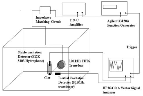
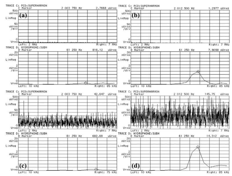
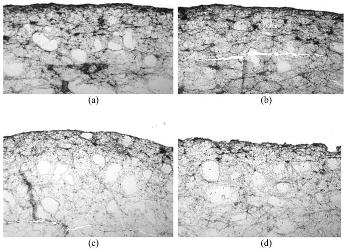
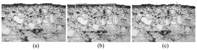
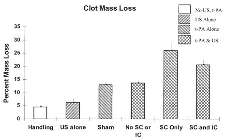
Pulsed ultrasound, when used as an adjuvant to recombinant tissue plasminogen activator (rt-PA), has been shown to enhance thrombolysis in the laboratory as well as in clinical trials for the treatment of ischemic stroke. The exact mechanism of this enhancement has not yet been elucidated. In this work, stable and inertial cavitation (SC and IC) are investigated as possible mechanisms for this enhancement. A passive cavitation detection scheme was utilized to measure cavitation thresholds at 120 kHz (80% duty cycle, 1667 Hz pulse repetition frequency) for four host fluid and sample combinations: plasma, plasma with rt-PA, plasma with clot and plasma with clot and rt-PA. Following cavitation threshold determination, clots were exposed to pulsed ultrasound for 30 min in vitro using three separate ultrasound treatment regimes: (1) no cavitation (0.15 MPa), (2) SC alone (0.24 MPa) or (3) SC + IC combined (0.36 MPa) in the presence of rt-PA. Percent clot mass loss after each treatment was used to determine thrombolysis efficacy. The highest percent mass loss was observed in the stable cavitation regime (26%), followed by the combined stable and inertial cavitation regime (20.7%). Interestingly, the percent mass loss in clots exposed to ultrasound without cavitation (13.7%) was not statistically significantly different from rt-PA alone (13%) [p > 0.05]. Significant enhancement of thrombolysis correlates with presence of cavitation and stable cavitation appears to play a more important role in the enhancement of thrombolysis. (E-mail: ).


Figures

Fig. 1
Fig. 1
Experimental apparatus showing the confocally aligned 120-kHz UTS transducer, stable cavitation detector and inertial cavitation detector, along with the electronic equipment used for cavitation detection. The water in the tank was kept at 37°C during all experiments.
Fig. 2
Fig. 2
Representative frequency spectrum traces from the HP89410A vector signal analyzer used for cavitation detection caused by exposure to 120-kHz ultrasound. The top trace in each panel is the frequency spectrum from 2 MHz to 7 MHz and the bottom trace is the frequency spectrum from 10 kHz to 75 kHz. (a) Shows the typical background noise level for plasma alone at 0.24 MPa P- when no cavitation is present. (b) Shows stable cavitation in plasma exposed to 0.36 MPa P- pressure amplitude. Note the 60-kHz peak on bottom trace corresponding to subharmonic emissions, but no broadband superharmonic emissions on the top trace typical of inertial cavitation. (c) Shows inertial cavitation in plasma with a clot exposed to 0.26 MPa P- pressure amplitude. Note the broadband superharmonic emissions corresponding to inertial cavitation. (d) Shows both inertial and stable cavitation present in plasma exposed to 0.40 MPa P-.
Fig. 3
Fig. 3
Immunohistochemical imaging analysis of treated clots. Following treatment, clots were fixed in formalin, dehydrated, embedded in paraffin, and cut in 4 micron sections. Clot slices were labeled with mouse antifibrinogen antibody and stained with horseradish peroxidase-linked goat antimouse IgG. Clot surfaces were photographed at 20 × magnification. All treated clots demonstrate a relatively densely staining band along the outermost surface. Relative to (a) Control, (b) The surface of the clot treated with ultrasound alone is essentially no different. (c) After treatment with rt-PA alone, the densely stained band at the surface is much thinner, but remains smooth in contour. (d) After treatment with rt-PA and ultrasound at 0.24 MPa P- (stable cavitation regime), the surface appears markedly irregular and porous.
Fig. 4
Fig. 4
Representative areas taken for mean gray-scale analysis to evaluate fibrin degradation. (a) Interior of the clot, (b) Surface and interior of the clot and (c) Surface of the clot.
Fig. 5
Fig. 5
Percent clot mass loss after 30 min exposure. n = 5 for each treatment protocol. Error bars indicate one standard deviation. “Handling” refers to clots in plasma alone (without rt-PA or pulsed ultrasound). “US alone” refers to clots without rt-PA, but with pulsed ultrasound (0.36 MPa P-), “Sham” refers to clots exposed to rt-PA alone, but without pulsed ultrasound. “No SC or IC” refers to clots exposed to rt-PA and pulsed ultrasound below both the stable cavitation (SC) and inertial cavitation (IC) thresholds (0.15 MPa P-). “SC Only” refers to clots with rt-PA and pulsed ultrasound above the stable but below the inertial cavitation threshold (0.24 MPa P-), “SC and IC” refers to clots exposed to rt-PA and pulsed ultrasound above the stable and inertial cavitation thresholds (0.36 MPa P-).
Fig. 6
Fig. 6
Immunohistochemical analysis of the effects of cavitation on treated clots. Clots were exposed to (a) rt-PA and ultrasound without cavitation, (b) With stable cavitation or (c) With stable and inertial cavitation for 30 min. Following treatment, the clots were prepared for immunohistochemical analysis. After treatment without any cavitation (a), the clot surface has a slightly irregular contour, but remains essentially smooth and intact. After treatment with stable cavitation but without inertial cavitation (b), the clot surface is notably irregular and discontinuous. After treatment with both stable and inertial cavitation (c), the clot surface appears thin but relatively smooth and continuous, similar in appearance to the clot treated with rt-PA alone (see Fig. 3c).

Similar articles

Cited by

References

    1. Akiyama M, Ishibashi T, Yamada T, Furuhata H. Low frequency ultrasound penetrates the cranium and enhances thrombolysis in vitro. Neurosurgery. 1998;43:828–833. - PubMed
    1. Alexandrov AV, Molina CA, Grotta JC, et al. Ultrasound-enhanced systemic thrombolysis for acute ischemic stroke. N Engl J Med. 2004;351(21):2170–2178. - PubMed
    1. Alexandrov AV, Demchul AM, Burgin WS, et al. Ultrasound-enhanced thrombolysis for acute ischemic stroke: Phase I. Findings of the CLOTBUST trial. J Neuroimaging. 2004;14(2):113–117. - PubMed
    1. ANSI Technical Report—Bubble detection and Cavitation Monitoring. ANSI. S1.24:TR-2002.
    1. Apfel RE. Acoustic cavitation prediction. J Acoust Soc Am. 1981;69:1624–1633.

Publication types