Review
. 2006 Nov;17(11):1481-1489.
doi: 10.1016/j.jasms.2006.06.006. Epub 2006 Jul 28.

Hydrogen exchange and mass spectrometry: A historical perspective

Affiliations
Review

Hydrogen exchange and mass spectrometry: A historical perspective

S Walter Englander. J Am Soc Mass Spectrom. 2006 Nov.

Abstract

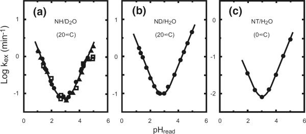
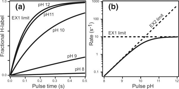
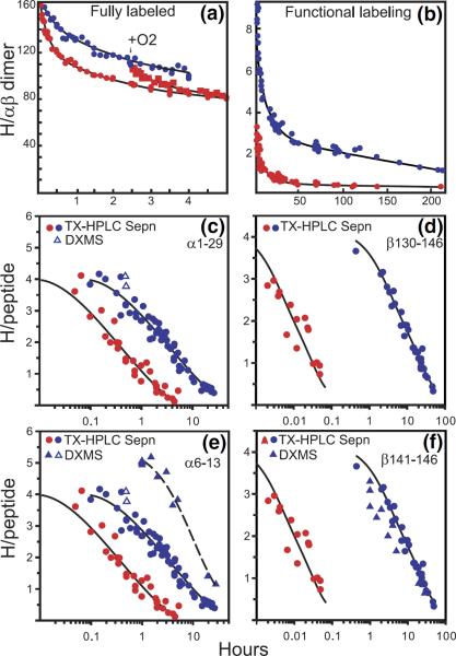
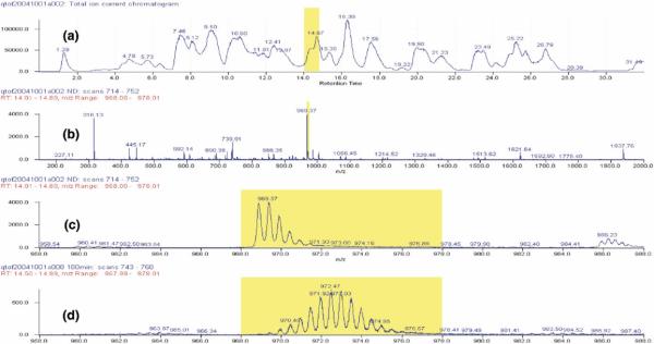
Protein molecules naturally emit streams of information-rich signals in the language of hydrogen exchange concerning the intimate details of their stability, dynamics, function, changes therein, and effects thereon, all resolved to the level of their individual amino acids. The effort to measure protein hydrogen exchange behavior, understand the underlying chemistry and structural physics of hydrogen exchange processes, and use this information to learn about protein properties and function has continued for 50 years. Recent work uses mass spectrometric analysis together with an earlier proteolytic fragmentation method to extend the hydrogen exchange capability to large biologically interesting proteins. This article briefly reviews the advances that have led us to this point and the understanding that has so far been achieved.


Figures

Figure 1
Figure 1
HX of the amide group in unstructured polypeptides. At the chemical level, amide HX rates vary linearly with hydronium and hydroxide ion concentration (eq 1). They depend also on temperature, neighboring side chains, and isotope effects [5, 6] (see URL HX2.Med.UPenn.edu/download). The HX halftime for unprotected amides is over 1 h at the pH minimum, between pH 2 and 3 at 0 °C, which makes possible the peptide separation/mass spectrometry technology.
Figure 2
Figure 2
HX of protected amides. The additional protection afforded by H-bonded structure depends on the thermodynamic and kinetic parameters of transient opening reactions that separate the H-bond and expose the hydrogen to solvent. Exchange rate increases linearly with solvent catalyst concentration in the EX2 range but will ultimately limit at the structural opening rate (EX1 limit). In the EX2 region, the reference chemical rate (eq 1, Figure 1) is decreased by the fraction of time that the hydrogen is exposed, given by kop/(kop + kcl) ≈ Kop (from eq 2), therefore can indicate the free-energy of the responsible structural opening reaction (eq 3). In the EX1 region, the HX rate becomes equal to, and therefore provides a way to measure, the rate for the responsible structural opening (eq 4). EX1 behavior can be documented by its insensitivity to pH as shown here or by correlated MS behavior when multiple amides experience the same EX1 exchange rate so that MS results show a molecular species moving from the unlabeled mass to the fully labeled mass in one concerted step.
Figure 3
Figure 3
HX results for hemoglobin (Hb) illustrating the progression of HX technology. (a) Whole Hb initially labeled to exchange equilibrium in tritiated water, then exchanged-out in oxy (red) and deoxy (blue) forms, measured by the early tritiumgel-filtration method [36]. Some allosterically sensitive sites are faster in oxyHb. When O2 is added to exchanging deoxy Hb, the sensitive sites are made faster and the separate curves merge. (b) Functional labeling results for whole Hb with exchange-in for only 1 min (pH 7.4, 0 °C [37]). The functional labeling method selectively labels some allosterically sensitive sites, making it possible to characterize their number and HX rates in both forms, and therefore the allosteric free-energy that they carry. (c) and (d) HX of functionally labeled Hb (35 min, 0 °C exchange-in) measured by the proteolytic fragmentation method with peptide separation by HPLC alone [38]. Two sets of allosterically sensitive amides are located at the segment level. They exchange by way of a local unfolding mechanism. (e) and (f) The integration of these approaches with mass spectrometric analysis which more closely defines some allosterically sensitive positions. (e) and (f) include MS data obtained with the same exchange-in as for the tritium experiments (open triangles), and more intensively (20 min, 20 °C, closed triangles) [35].
Figure 4
Figure 4
H-D exchange/MS experiment. Amyloid (α-synuclein) HX labeled by timed exposure to D2O was dissociated with chemical denaturant at the condition of minimum HX rate (Figure 1), then passed through two immobilized acid protease columns, the proteolytic peptides were roughly resolved by HPLC, and then by on line ESI MS. (a) TIC trace of the HPLC eluant. (b) HPLC eluant was continually scanned by ESI MS, as shown for the eluant region marked in (a) MS results for the peptide ion marked in (b) are shown expanded for the protonated (c) and the partially deuterated (d) condition. Subtraction of the centroid mass of the unlabelled all-H peptide from the D-labeled peptide yields the number of D-labeled sites recovered on each peptide.
Figure 5
Figure 5
H-D exchange/MS results for allosteric structure change in hemoglobin obtained as in Figure 4. Some allosterically sensitive amide sites were initially selectively labeled by the functional labeling method. Exchange-out results are compared for R-state oxy (red) and T-state deoxy (blue) Hb (obtained by subtracting the background from the raw data, both shown in gray; experimental details in [35]). HX was measured by the proteolytic fragmentation-separation method with analysis by MS. The peptides indicated span the entire length of the α- and the β-chains. The results show where changes occur and where they do not, and evaluate the number of amide sites affected within each sensitive segment. Several of the measured changes strongly suggest multi-residue segmental unfolding reactions (local unfolding) in which case the change in structural free-energy deposited at each allosterically sensitive position can be read out in terms of the change in HX rate between the two different allosteric forms (eq 3).
Figure 6
Figure 6
H-D exchange/MS results for α-synuclein obtained as in Figure 4. Results for the soluble α-synuclein monomer (green) compare well with expected unprotected rates (dashed black curves). Results for the insoluble amyloid, shown in red, identify the well-protected chain segment that participates in the amyloid cross-β region and show the degree of protection in other regions.

References

    1. Hvidt A, Nielsen SO. Hydrogen exchange in proteins. Adv. Protein Chem. 1966;21:287–386. - PubMed
    1. Eigen M. Proton transfer, acid-base catalysis, and enzymatic hydrolysis. Angew. Chem. Int. Ed. English. 1964;3:1–19.
    1. Englander SW, Kallenbach NR. Hydrogen exchange and structural dynamics of proteins and nucleic-acids. Q. Rev. Biophys. 1984;16:521–655. - PubMed
    1. Perrin CL. Proton exchange in amides: Surprises from simple systems. Accts. Chem. Res. 1989;22:268–275.
    1. Bai Y, Milne JS, Mayne L, Englander SW. Primary structure effects on peptide group hydrogen exchange. Proteins. 1993;17:75–86. - PMC - PubMed

Publication types