. 2006 Oct 10;354(1):132-42.
doi: 10.1016/j.virol.2006.06.026. Epub 2006 Jul 31.

The human severe acute respiratory syndrome coronavirus (SARS-CoV) 8b protein is distinct from its counterpart in animal SARS-CoV and down-regulates the expression of the envelope protein in infected cells

Affiliations

The human severe acute respiratory syndrome coronavirus (SARS-CoV) 8b protein is distinct from its counterpart in animal SARS-CoV and down-regulates the expression of the envelope protein in infected cells

Choong-Tat Keng et al. Virology. .

Abstract

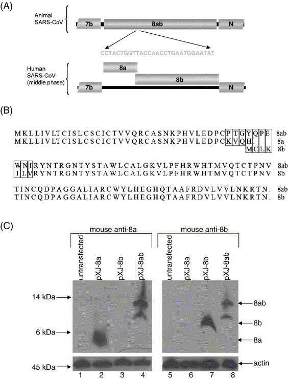
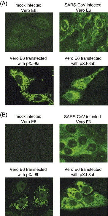
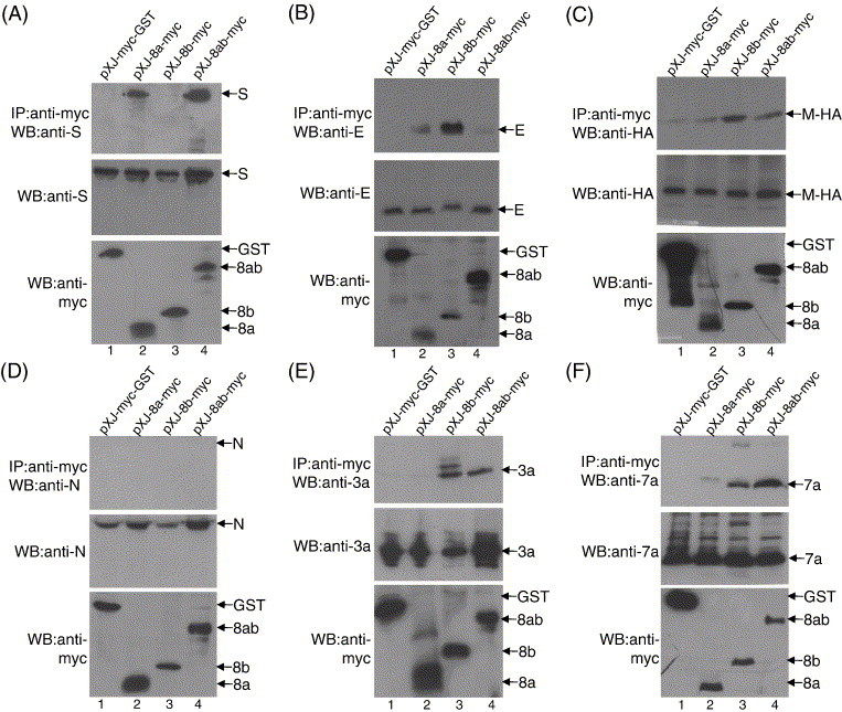
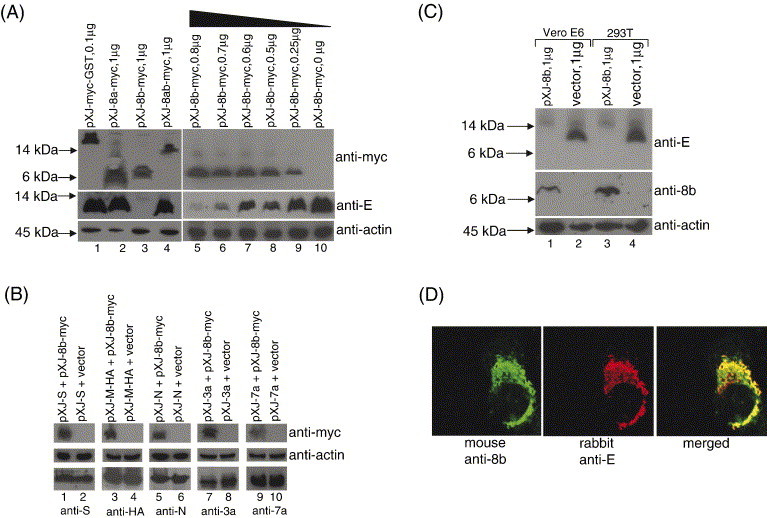
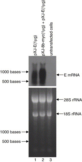
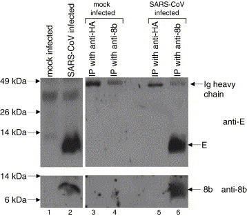
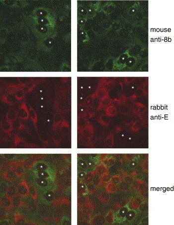
The severe acute respiratory syndrome coronavirus (SARS-CoV), isolated from humans infected during the peak of epidemic, encodes two accessory proteins termed as 8a and 8b. Interestingly, the SARS-CoV isolated from animals contains an extra 29-nucleotide in this region such that these proteins are fused to become a single protein, 8ab. Here, we compared the cellular properties of the 8a, 8b and 8ab proteins by examining their cellular localizations and their abilities to interact with other SARS-CoV proteins. These results may suggest that the conformations of 8a and 8b are different from 8ab although nearly all the amino acids in 8a and 8b are found in 8ab. In addition, the expression of the structural protein, envelope (E), was down-regulated by 8b but not 8a or 8ab. Consequently, E was not detectable in SARS-CoV-infected cells that were expressing high levels of 8b. These findings suggest that 8b may modulate viral replication and/or pathogenesis.


Figures

Fig. 1
Fig. 1
Expressions of SARS-CoV 8a, 8b and 8ab proteins. (A) Schematic diagram showing the genetic differences in the ORF8 region of the SARS-CoV isolated from animals and humans infected during the middle phase of the SARS epidemic in 2003 (modified from Guan et al., 2003). The animal isolates have an extra 29-nucleotides insertion such that the subgenomic RNA encodes for a single protein, termed 8ab, whereas that of the human isolates (from the middle phase) encodes two proteins, 8a and 8b. Human isolates from early and late phases of the epidemic also have the 29-nucleotides insertion found in the animal SARS-CoV. (B) Alignment of the sequences of 8a, 8b and 8ab proteins used in this study. Mismatches between 8a and 8ab or 8b and 8ab are boxed. The 8ab is reconstructed from a human isolate from the middle phase (SIN2774) by insertion of the 29-nucletoides found in a human isolate from the early phase (GZ02). (C) Western blot analysis was performed to detect 8a, 8b and 8ab proteins expressed in Vero E6 cells using cDNA constructs. The experiments were performed with either mouse anti-8a polyclonal antibody (upper panel, lanes 1–4) or mouse anti-8b polyclonal antibody (upper panel, lanes 5–8). Equal amounts of cells were used in each lane as verified by the level of endogenous actin (bottom panel).
Fig. 2
Fig. 2
Cellular localizations of 8a and 8b in SARS-CoV-infected cells and Vero E6 cells transfected with DNA constructs for expressing 8a, 8b and 8ab. Specific mouse anti-8a and anti-8b polyclonal antibodies were used in indirect immunofluorescence experiments to determine the expressions of (A) 8a and (B) 8b, respectively. The top two panels showed the specific reactivities of the anti-8a and anti-8b antibodies to proteins expressed in SARS-CoV-infected cells (right panels) as no unspecific staining was observed for the mock-infected cells (left panels). The bottom two panels showed the reactivities of the antibodies to 8a and 8ab (A) or 8b and 8ab (B) expressed in Vero E6 by transfection of cDNA constructs.
Fig. 3
Fig. 3
Interactions of 8a, 8b and 8ab with other SARS-CoV proteins. Cell lysates containing myc-GST (lane 1), 8a-myc (lane 2), 8b-myc (lane 3) or 8ab-myc (lane 4) and another SARS-CoV protein (S, E, M-HA, N, 3a or 7a) were immunoprecipitated with an anti-myc polyclonal antibody and protein A-agarose beads. (A) The amounts of S protein co-immunoprecipitated (IP) by the myc-tagged proteins were determined using an anti-S monoclonal antibody (top panel). The amounts of S and myc-tag proteins in the lysates before co-immunoprecipitation were also determined by Western blot (WB) with anti-S and anti-myc monoclonal antibodies, respectively (middle and bottom panels). The same experiments were performed for panels B–F except that different antibodies against the specific viral proteins were used, namely (B) anti-E mouse polyclonal; (C) anti-HA monoclonal (as the M protein fused with a HA tag at the C terminus); (D) anti-N mouse polyclonal; (E) anti-3a mouse polyclonal; (F) anti-7a mouse polyclonal.
Fig. 4
Fig. 4
Effects of 8b on the expression of the small structural protein, E. (A) 293T cells were co-transfected with 2 μg of pXJ-E and 0.1 μg of pXJ-myc-GST (lane 1), 1 μg of pXJ-8a-myc (lane 2), pXJ-8b-myc (lane 3), pXJ-8ab-myc (lane 4), or decreasing amount of pXJ-8b-myc (lanes 5–10). Total cell lysates were subjected to Western blot analysis to determine the expression of E (middle panel) and myc-tagged proteins (top panel). Equal amounts of cells were used in each lane as verified by the level of endogenous actin (bottom panel). (B) 293T cells were co-transfected with 1 μg of pXJ-S and 1 μg of either pXJ-8b-myc or empty vector (lanes 1 and 2). Total cell lysates were subjected to Western blot analysis to determine the expression of 8b-myc (top panel) and S (lower panel). Equal amounts of cells were used in each lane as verified by the level of endogenous actin (middle panel). Similar experiments were performed with 1 μg of pXJ-M-HA (lanes 3 and 4), 0.25 μg of pXJ-N (lanes 5 and 6), 0.4 μg of pXJ-3a (lanes 7 and 8) or 0.4 μg of pXJ-7a (lanes 9 and 10). (C) Vero E6 or 293T cells were co-transfected with 2 μg of pXJ-E and 1 μg of pXJ-8b (lanes 1 and 3) or 1 μg of empty vector (lanes 2 and 4). Total cell lysates were subjected to Western blot analysis to determine the expression of E (top panel) and 8b (middle panel). Equal amounts of cells were used in each lane as verified by the level of endogenous actin (bottom panel). (D) Indirect immunofluorescence experiments were performed to determine the cellular localization of 8b and E in Vero E6 cells co-transfected with pXJ-8b and pXJ-E. The expression of 8b is represented by FITC staining (left panel), whereas the expression of E is represented by rhodamine staining (middle panel). The merged images showed that the 8b and E partially colocalized in co-transfected cells (right panel).
Fig. 5
Fig. 5
Effects of 8b protein on the transcription of the E gene determined by Northern blot analysis. Equal amount of total RNA (15 μg) isolated from 293T cells transfected with pXJ-E (lane 1), pXJ-8b-myc and pXJ-E (lane 2) cDNA constructs or untransfected 293T (lane 3) was separated on a denaturing agarose gel and transferred to nylon membrane. The amounts of E mRNA present were determined by hybridization with an E gene-specific probe (top panel). In order to verify that equal amounts of total RNA were loaded in each lane of the agarose gel before transfer, the amounts of 18S and 28S ribosomal RNA were visualized under UV light (bottom panel).
Fig. 6
Fig. 6
Interaction between E and 8b in SARS-CoV-infected cells determined by co-immunoprecipitation experiment. Lysates from mock-infected or SARS-CoV-infected cells were immunoprecipitated using rabbit anti-8b polyclonal antibody (lanes 4 and 6) or an irrelevant antibody (rabbit anti-HA polyclonal antibody, lanes 3 and 5) and protein A-agarose beads. Western blot analyses were then performed to determine the amount of E (upper panel) or 8b (lower panel) present in the lysates before immunoprecipitation (lanes 1 and 2) and the immunocomplexes on the protein A-agarose beads (lanes 3–6).
Fig. 7
Fig. 7
Expressions of E and 8b in SARS-CoV-infected cells determined by indirect immunofluorescence experiments. The expression of 8b was represented by FITC staining (top row), whereas the expression of E was represented by Rhodamine staining (middle row). The merged images showed that the expression of 8b and E were mutually exclusive (bottom row). Two representative sets of data were presented and cells expressing high levels of 8b were marked with white asterisks.

Similar articles

Cited by

References

    1. Chiu R.W., Chim S.S., Tong Y.K., Fung K.S., Chan P.K., Zhao G.P., Lo Y.M. Tracing SARS-coronavirus variant with large genomic deletion. Emerging Infect. Dis. 2005;11:168–170. - PMC - PubMed
    1. Donnelly C.A., Fisher M.C., Fraser C., Ghani A.C., Riley S., Ferguson N.M., Anderson R.M. Epidemiological and genetic analysis of severe acute respiratory syndrome. Lancet Infect. Dis. 2004;4:672–683. - PMC - PubMed
    1. Drosten C., Preiser W., Gunther S., Schmitz H., Doerr H.W. Severe acute respiratory syndrome: identification of the etiological agent. Trends Mol. Med. 2003;9:325–327. - PMC - PubMed
    1. Fielding B.C., Tan Y.J., Shuo S., Tan T.H., Ooi E.E., Lim S.G., Ho W., Goh P.Y. Characterization of a unique group-specific protein (U122) of the severe acute respiratory syndrome (SARS) coronavirus. J. Virol. 2004;78:7311–7318. - PMC - PubMed
    1. Guan Y., Zheng B.J., He Y.Q., Liu X.L., Zhuang Z.X., Cheung C.L., Luo S.W., Li P.H., Zhang L.J., Guan Y.J., Butt K.M., Wong K.L., Chan K.W., Lim W., Shortridge K.F., Yuen K.Y., Peiris J.S., Poon L.L. Isolation and characterization of viruses related to the SARS coronavirus from animals in southern China. Science. 2003;302:276–278. - PubMed

Publication types

MeSH terms

LinkOut - more resources