. 2006 Aug;169(2):338-46.
doi: 10.2353/ajpath.2006.060312.

Hematopoietic stem cells: the paradigmatic tissue-specific stem cell

Affiliations

Hematopoietic stem cells: the paradigmatic tissue-specific stem cell

David Bryder et al. Am J Pathol. 2006 Aug.

Erratum in

  • Am J Pathol. 2006 Nov;169(5):1899

Abstract

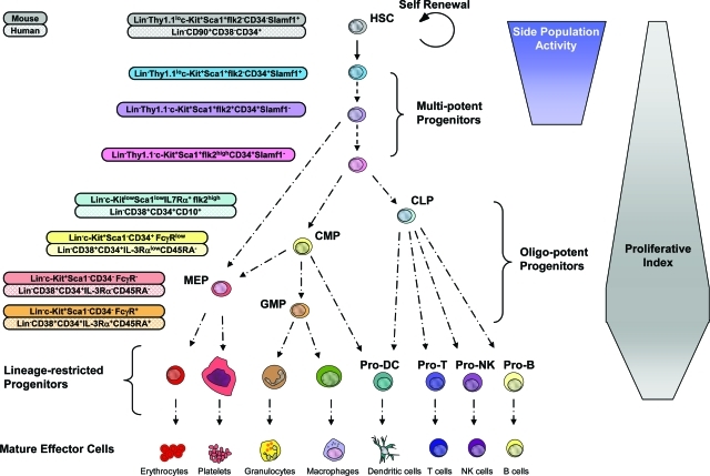
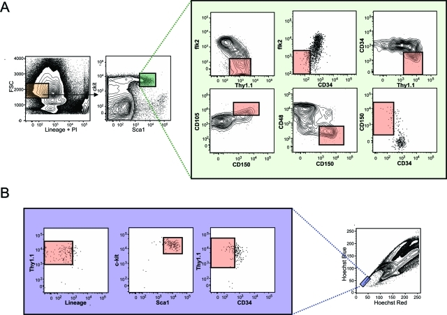
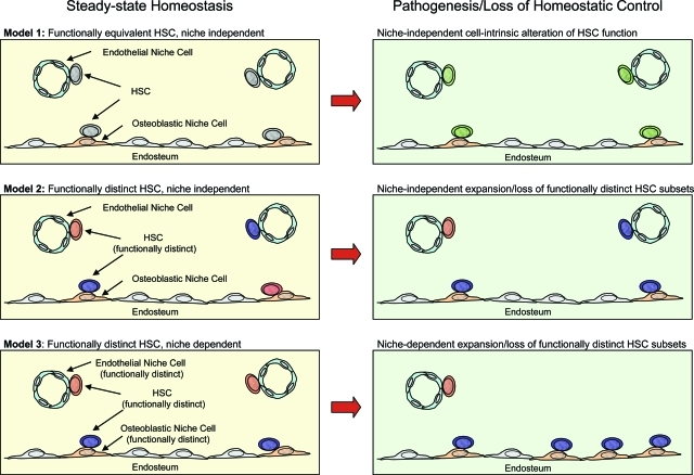
The recent prospective isolation of a wide variety of somatically derived stem cells has affirmed the notion that homeostatic maintenance of most tissues and organs is mediated by tissue-specific stem and progenitor cells and fueled enthusiasm for the use of such cells in strategies aimed at repairing or replacing damaged, diseased, or genetically deficient tissues and organs. Hematopoietic stem cells (HSCs) are arguably the most well-characterized tissue-specific stem cell, with decades of basic research and clinical application providing not only a profound understanding of the principles of stem cell biology, but also of its potential pitfalls. It is our belief that emerging stem cell fields can benefit greatly from an understanding of the lessons learned from the study of HSCs. In this review we discuss some general concepts regarding stem cell biology learned from the study of HSCs with a highlight on recent work pertaining to emerging topics of interest for stem cell biology.


Figures

Figure 1-6946
Figure 1-6946
Model of the hematopoietic developmental hierarchy. Self-renewing HSCs reside at the top of the hierarchy, giving rise to a number of multipotent progenitors. Multipotent progenitors give rise to oligo-potent progenitors including the CLP, which gives rise to mature B lymphocytes, T lymphocytes, and natural killer (NK) cells. The common myeloid progenitor (CMP) gives rise to granulocyte-macrophage progenitors, which differentiate into monocytes/macrophages and granulocytes, and megakaryocyte/erythrocyte progenitors, which differentiate into megakaryocytes/platelets and erythrocytes. Both CMPs and CLPs have been proposed to give rise to dendritic cells. The cell surface phenotype of many of these cell types is shown for the murine and human systems. It should be noted that certain markers such as Thy1.1 and ScaI are only informative in some but not all mouse strains. Development from the oligo-potent progenitors to mature blood cells proceeds through a number of intermediate progenitors (not shown). It should be noted that the absolute lineage potential and developmental relationships of some of the subsets indicated have not yet been fully resolved. For example, evidence suggests that the megakaryocyte and erythrocyte lineages may originate from a multipotent progenitor and not pass through a CMP intermediate. The developmental passage of HSCs through multipotent progenitors, oligo-potent progenitors, and lineage-specific progenitors is generally associated with increases in proliferative index although this trend is not absolute and has not been resolved for all stages of development. The capacity to efflux dyes such as Hoechst 33342 or rhodamine-123 (termed side population activity) is restricted to HSCs and its immediate multipotent progenitors. It should be noted that although the different multipotent progenitor subsets in mice have been functionally resolved to a significant degree, this is not true of the human system. We therefore believe that cells defined as human HSCs by the cell surface markers LinCD90+CD38CD34+ are likely to include one or more multipotent progenitor populations.
Figure 2-6946
Figure 2-6946
Heterogeneity within the primitive hematopoietic compartment. A: Cell surface staining of the murine KLS compartment with markers that enrich for stem cell activity. In bone marrow, all HSC activity is found within the lineage-negative (orange box: negative for antigens found on mature blood cells including B220, Mac1, Gr-1, Il7Rα, Ter119, CD3, CD4, CD8) and ScaIhigh and c-kithigh fractions (small green box). Because only ∼1 in 30 cells in the KLS compartment is a long-term multilineage reconstituting HSC, additional cell surface markers are used to enrich for HSC activity as illustrated in the expanded green box. These include positive cell surface markers such as Thy1.1, CD105 (Endoglin), and CD150 (Slamf1), in addition to negative markers including CD34, CD48, and flk2. In each fluorescence-activated cell sorting plot, the pink box denotes where HSC activity is found as determined by transplantation experiments performed by others and ourselves. It should be noted that the staining combinations shown only represent enrichment strategies, and combinations of numerous markers are recommended to further enrich for HSC activity. For example, although flk2 negativity and Thy1.1 positivity enrich for HSC activity from the KLS compartment, the addition of CD34 negativity adds additional significant enrichment of HSC activity. It should also be noted that Thy1.1 expression is only useful as a marker on HSCs if using mouse strains carrying the Thy1.1 allele, and selection based on negativity for CD34 requires the use of mice of more than 8 weeks of age. B: Cell surface marker distribution within the fraction of bone marrow cells exhibiting the highest ability to efflux Hoechst 33342 (side population tip cells). HSC activity is only found within the pink boxes of each plot as determined by transplantation experiments performed by others and ourselves.
Figure 3-6946
Figure 3-6946
Modeling HSC functional capacity in the context of divergent stem cell niches and pathogenesis. Three models of HSC/niche functional interaction are depicted under normal steady-state homeostatic conditions (left) and pathogenic conditions, or conditions in which loss of homeostatic control is manifested at the stem cell level (right). In model 1 (top left), all HSCs have the same functional capacity regardless of association with osteoblastic or endothelial niche cells. Pathogenic change at the stem cell level is cell intrinsic in this model (top right) and is independent of divergent niche interactions. In model 2 (middle left), functionally distinct HSCs exist (greater or lesser lineage potential, self-renewal, homing capacity, and so forth), and their functional identity is cell intrinsic and independent of interactions with divergent niches. Stem cell pathogenic change in this model (middle right) may be characterized by the selective expansion or loss of distinct HSC subsets independent of divergent niche interactions. In model 3 (bottom left), the functional identity of HSCs is dependent on interactions with divergent microenvironments and is therefore not cell intrinsic. Stem cell pathogenic change in this model (bottom right) is characterized by the selective expansion or loss of specific niche subtypes.

References

    1. Uchida N, Buck DW, He D, Reitsma MJ, Masek M, Phan TV, Tsukamoto AS, Gage FH, Weissman IL. Direct isolation of human central nervous system stem cells. Proc Natl Acad Sci USA. 2000;97:14720–14725. - PMC - PubMed
    1. Sherwood RI, Christensen JL, Conboy IM, Conboy MJ, Rando TA, Weissman IL, Wagers AJ. Isolation of adult mouse myogenic progenitors: functional heterogeneity of cells within and engrafting skeletal muscle. Cell. 2004;119:543–554. - PubMed
    1. Blanpain C, Lowry WE, Geoghegan A, Polak L, Fuchs E. Self-renewal, multipotency, and the existence of two cell populations within an epithelial stem cell niche. Cell. 2004;118:635–648. - PubMed
    1. Stingl J, Eirew P, Ricketson I, Shackleton M, Vaillant F, Choi D, Li HI, Eaves CJ. Purification and unique properties of mammary epithelial stem cells. Nature. 2006;439:993–997. - PubMed
    1. Morrison SJ, White PM, Zock C, Anderson DJ. Prospective identification, isolation by flow cytometry, and in vivo self-renewal of multipotent mammalian neural crest stem cells. Cell. 1999;96:737–749. - PubMed