. 2006 Aug;169(2):388-99.
doi: 10.2353/ajpath.2006.050964.

Nitric oxide is an important mediator of renal tubular epithelial cell death in vitro and in murine experimental hydronephrosis

Affiliations

Nitric oxide is an important mediator of renal tubular epithelial cell death in vitro and in murine experimental hydronephrosis

Tiina Kipari et al. Am J Pathol. 2006 Aug.

Abstract

Macrophages play a pivotal role in tissue injury and fibrosis during renal inflammation. Although macrophages may induce apoptosis of renal tubular epithelial cells, the mechanisms involved are unclear. We used a microscopically quantifiable co-culture assay to dissect the cytotoxic interaction between murine bone marrow-derived macrophages and Madin-Darby canine kidney cells and primary murine renal tubular epithelial cells. The induction of tubular cell apoptosis by cytokine-activated macrophages was reduced by inhibitors of nitric oxide synthase whereas tubular cell proliferation was unaffected. Furthermore, cytokine-activated macrophages derived from mice targeted for the deletion of inducible nitric oxide synthase were noncytotoxic. We then examined the role of nitric oxide in vivo by inhibiting inducible nitric oxide synthase in the model of murine experimental hydronephrosis. l-N(6)-(1-iminoethyl)-lysine was administered in the drinking water between days 5 and 7 after ureteric obstruction. Macrophage infiltration was comparable between groups, but treatment significantly inhibited tubular cell apoptosis at day 7. Tubular cell proliferation was unaffected. Inducible nitric oxide synthase blockade also reduced interstitial cell apoptosis and increased collagen III deposition. These data indicate that nitric oxide is a key mediator of macrophage-directed tubular cell apoptosis in vitro and in vivo and also modulates tubulointerstitial fibrosis.


Figures

Figure 1-6924
Figure 1-6924
Cytokine-activated macrophages induce MDCK cell apoptosis. A: MDCK cells were cultured alone or in the presence of mature murine bone marrow-derived macrophages in the presence or absence of LPS and IFN-γ. After a 24-hour incubation the level of MDCK cell apoptosis is determined by in situ fixation of cultures with formaldehyde and fluorescence microscopy after staining with Hoechst. Cytokine-activated macrophages induce significant MDCK cell death. *P < 0.001 versus nonactivated co-culture (data from experiments with macrophages from four different mice). B: Low-power view of cytokine-activated co-culture of macrophages (unlabeled) and CellTracker green-labeled MDCK cells after 24 hours. Apoptotic MDCK cells are visible as bright cells exhibiting cytoplasmic condensation. C: The bright rounded apoptotic MDCK cell exhibits nuclear pyknosis (green arrow); a classical feature of apoptosis. The nucleus of an adjacent unlabeled macrophage is also evident (red arrow). The merged image demonstrates the proximity of the macrophage to the MDCK cell.
Figure 2-6924
Figure 2-6924
Macrophage-derived NO is an important mediator of MDCK cell apoptosis but is not involved in the inhibition of MDCK cell proliferation. A: MDCK cells were cultured alone or in the presence of bone marrow-derived macrophages derived from either iNOS wild-type (WT) or knockout (KO) mice. Cultures were activated with LPS and IFN-γ in the presence or absence of the NOS inhibitor L-NAME (200 μmol/L) or control D-NAME (200 μmol/L). iNOS knockout macrophages are not cytotoxic under any conditions whereas the cytotoxicity of iNOS wild-type macrophages is completely abrogated by pharmacological inhibition of NO production. *P < 0.05 versus nonactivated iNOS wild-type macrophage co-cultures (data from experiments with macrophages from three different mice). B: MDCK cells were cultured alone or in the presence of mature bone marrow-derived macrophages derived from either iNOS wild-type or knockout mice. Cultures were activated with LPS and IFN-γ in the presence or absence of the NOS inhibitor L-NAME. Inhibition of MDCK cell proliferation by macrophages is independent of cytokine activation, macrophage genotype, or pharmacological inhibition of NO production. *P < 0.05 versus MDCK cells alone (data from experiments with macrophages from three different mice).
Figure 3-6924
Figure 3-6924
Cytokine-activated macrophages induce apoptosis in primary murine tubular epithelial cells (PTEs). A: PTE cells were cultured in the presence of mature murine bone marrow-derived macrophages. Selected cell cultures were activated with LPS and IFN-γ in the presence or absence of the NOS inhibitor L-NAME (200 μmol/L). *P < 0.05 versus activated co-cultures in the presence or absence of L-NAME (data from experiments with cells from three different mice). B: PTE cells were cultured in the presence of mature bone marrow-derived macrophages derived from either iNOS wild-type or knockout mice. Selected cell cultures were activated with LPS and IFN-γ. Cytokine activation of iNOS wild-type macrophages induced PTE cell apoptosis whereas activated iNOS knockout macrophages were noncytotoxic. *P < 0.05 versus nonactivated co-culture (data from experiments with cells from five different mice). C: Photomicrographs from a co-culture of fluorescently labeled macrophages (CellTracker orange) and PTE cells (CellTracker green) demonstrating evidence of macrophage recognition and phagocytosis of apoptotic tubular cells. The merged image demonstrates a condensed green apoptotic tubular cell with a pyknotic nucleus that has been ingested by a CellTracker orange-labeled macrophage.
Figure 4-6924
Figure 4-6924
Obstructed kidneys exhibit tubulointerstitial infiltration with macrophages and iNOS-positive cells. Tissue sections were immunostained for the macrophage marker F4/80 and/or iNOS. A: Normal nonmanipulated kidneys exhibit occasional F4/80-positive resident macrophages (arrow). B: In contrast, obstructed kidneys exhibit a prominent interstitial population of infiltrating F4/80-positive macrophages (arrow). C–E: Fluorescent photomicrographs showing double-immunofluorescent staining of iNOS and F4/80 in day 7 obstructed kidney: iNOS-positive cells stained green (C), F4/80-positive interstitial macrophages stained red (D), and merged image demonstrating co-localization (yellow) of iNOS and F4/80 indicating the presence of infiltrating macrophages expressing iNOS (E).
Figure 5-6924
Figure 5-6924
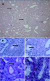
Obstructed kidneys exhibit apoptosis and proliferation of tubular epithelial and interstitial cells. Tissue sections underwent TUNEL staining to detect apoptotic cells whereas cells undergoing mitosis were identified on PAS-stained tissue sections. A: Low-power view of obstructed kidneys demonstrating scattered TUNEL-positive apoptotic cells (arrows). B: High-power view of a TUNEL-positive apoptotic tubular epithelial cell (arrow). C: High-power view of a TUNEL-positive apoptotic interstitial cell (arrow). D: High-power view of a tubular epithelial cell undergoing mitosis (arrow). E: High-power view of an interstitial cell undergoing mitosis (arrow). Original magnifications: ×200 (A); ×1000 (B–E).
Figure 6-6924
Figure 6-6924
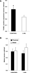
Pharmacological blockade of iNOS reduces tubular cell apoptosis but does not affect tubular cell proliferation after ureteric obstruction. Mice were administered L-NIL, an irreversible pharmacological inhibitor of the enzyme iNOS, or the isomeric control D-NIL, and the obstructed kidneys were removed at day 7. A: The level of tubular cell apoptosis in obstructed kidneys is significantly reduced after the administration of L-NIL. *P < 0.05 versus control (eight mice per group). B: The level of tubular cell proliferation in obstructed kidneys is unaffected after the administration of L-NIL (n = seven to eight mice per group).
Figure 7-6924
Figure 7-6924
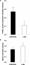
Pharmacological blockade of iNOS reduces interstitial cell apoptosis but does not affect interstitial cell proliferation after ureteric obstruction. Mice were administered L-NIL, an irreversible pharmacological inhibitor of the enzyme iNOS, or the isomeric control D-NIL, and the obstructed kidneys were removed at day 7. A: The level of interstitial cell apoptosis in obstructed kidneys is significantly reduced after the administration of L-NIL. *P < 0.05 versus control (eight mice per group). B: The level of interstitial cell proliferation in obstructed kidneys is unaffected after the administration of L-NIL (n = seven to eight mice per group).
Figure 8-6924
Figure 8-6924
Pharmacological blockade of iNOS increases tubulointerstitial deposition of collagen III after ureteric obstruction. Mice were administered L-NIL, an irreversible pharmacological inhibitor of the enzyme iNOS, or the isomeric control D-NIL, and the obstructed kidneys were removed at day 7. A: Photomicrographs of collagen III immunostaining at day 7 in control mice and mice treated with L-NIL. B: The administration of L-NIL increases the tubulointerstitial deposition of collagen III assessed by computer image analysis. *P < 0.05 versus control (n = seven to eight mice per group).

Similar articles

Cited by

References

    1. Gordon S. Macrophage-restricted molecules: role in differentiation and activation. Immunol Lett. 1999;65:5–8. - PubMed
    1. Leibovich SJ, Ross R. The role of the macrophage in wound repair A study with hydrocortisone and antimacrophage serum. Am J Pathol. 1975;78:71–100. - PMC - PubMed
    1. Gouon-Evans V, Rothenberg ME, Pollard JW. Postnatal mammary gland development requires macrophages and eosinophils. Development. 2000;127:2269–2282. - PubMed
    1. Lang RA, Bishop JM. Macrophages are required for cell death and tissue remodeling in the developing mouse eye. Cell. 1993;74:453–462. - PubMed
    1. Savill J, Dransfield I, Gregory C, Haslett C. A blast from the past: clearance of apoptotic cells regulates immune responses. Nat Rev Immunol. 2002;2:965–975. - PubMed

Publication types

MeSH terms

LinkOut - more resources