. 2006 Aug;169(2):405-15.
doi: 10.2353/ajpath.2006.060049.

Integrin-mediated transforming growth factor-beta activation regulates homeostasis of the pulmonary epithelial-mesenchymal trophic unit

Affiliations

Integrin-mediated transforming growth factor-beta activation regulates homeostasis of the pulmonary epithelial-mesenchymal trophic unit

Jun Araya et al. Am J Pathol. 2006 Aug.

Abstract

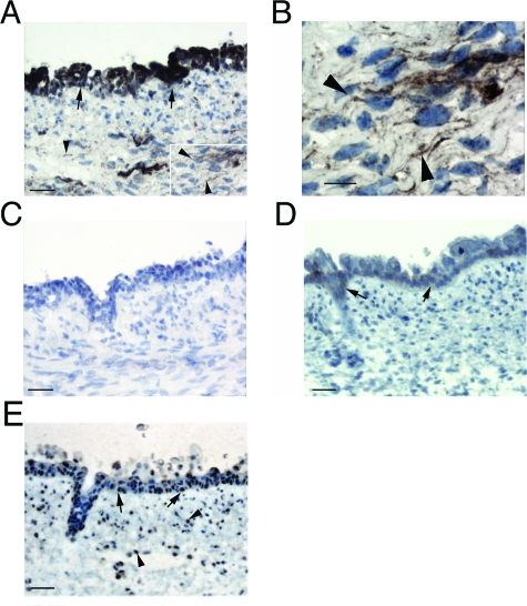
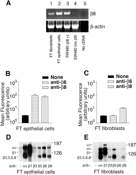
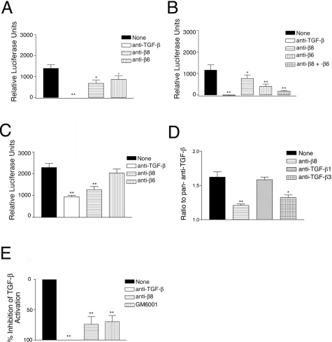
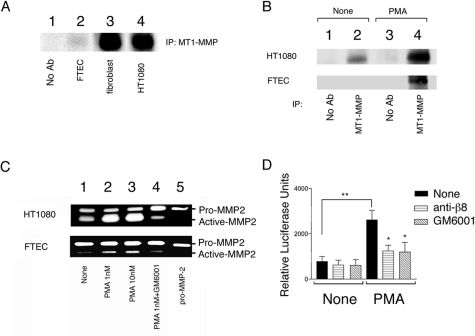
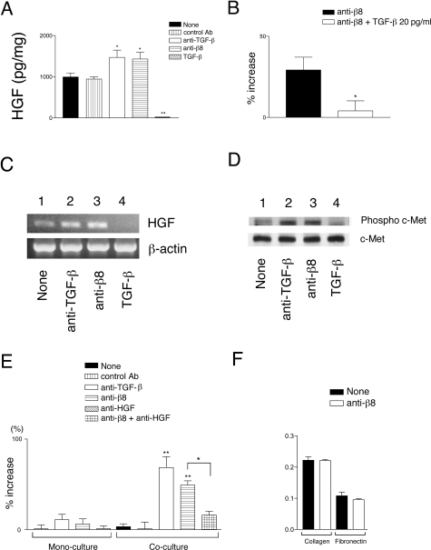
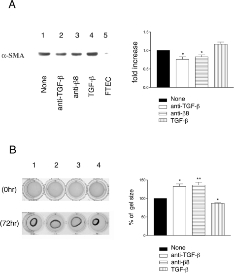
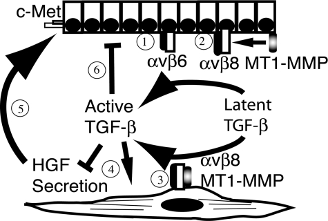
Trophic interactions between pulmonary epithelial and mesenchymal cell types, known as the epithelial-mesenchymal trophic unit (EMTU), are crucial in lung development and lung disease. Transforming growth factor (TGF)-beta is a key factor in mediating these interactions, but it is expressed in a latent form that requires activation to be functional. Using intact fetal tracheal tissue and primary cultures of fetal tracheal epithelial cells and fibroblasts, we demonstrate that a subset of integrins, alpha(v)beta(6) and alpha(v)beta(8), are responsible for almost all of the TGF-beta activation in the EMTU. Both alpha(v)beta(8) and alpha(v)beta(6) contribute to fetal tracheal epithelial activation of TGF-beta, whereas only alpha(v)beta(8) contributes to fetal tracheal fibroblast activation of TGF-beta. Interestingly, fetal tracheal epithelial alpha(v)beta(8)-mediated TGF-beta activation can be enhanced by phorbol esters, likely because of the increased activity of MT1-MMP, an essential co-factor in alpha(v)beta(8)-mediated activation of TGF-beta. Autocrine alpha(v)beta(8)-mediated TGF-beta activation by fetal tracheal fibroblasts results in suppression of both transcription and secretion of hepatocyte growth factor, which is sufficient to affect phosphorylation of the airway epithelial hepatocyte growth factor receptor, c-Met, as well as airway epithelial proliferation in a co-culture model of the EMTU. These findings elucidate the function and complex regulation of integrin-mediated activation of TGF-beta within the EMTU.


Figures

Figure 1-6929
Figure 1-6929
Integrin αvβ8 is expressed by fetal tracheal epithelial cells and fibroblasts. Immunohistochemistry of fetal tracheas was performed using integrin β8 (A–C) and β6 (D) subunit-specific or phosphorylated SMAD-2 antibodies (E). In A, inset indicates area magnified in B. In C, the primary anti-β8 antibody was preincubated with a GST-β8 cytoplasmic domain fusion protein. Epithelial and mesenchymal staining is indicated by arrows and arrowheads, respectively. Scale bars: 50 μm (A, CE); 10 μm (B).
Figure 2-6929
Figure 2-6929
The integrin αvβ8 is expressed by fetal tracheal epithelial cells and fibroblasts. A: RT-PCR for the β8 integrin subunit (top) and β-actin (bottom) was performed from total RNA harvested from fetal tracheal fibroblasts (lane 1), fetal tracheal epithelial cells (lane 2), wild-type SW480 cells stably transfected with a β8 expression construct (lane 3), SW480 cells (lane 4), and a control with no added cDNA (lane 5). Shown are bands migrating at the appropriate sizes for the amplification products. Flow cytometry of passage 1 fetal tracheal epithelial cells (B) and fetal tracheal fibroblasts (C) was performed using either no primary antibody (black bars), anti-β6 (horizontal hatched bars), or anti-β8 (vertical hatched bars). Results are expressed as the mean fluorescence intensity in arbitrary fluorescence units (log10). Immunoprecipitation of biotin surface-labeled fetal tracheal (FT) epithelial cells (D) or fetal tracheal fibroblasts (E) using no primary antibody or anti-integrin subunit and complex specific antibodies against αv (L230), β1 (P5D2), β3 (AP3), αvβ5 (P1F6), αvβ6 (E7P6), or αvβ8 (14E5). Antibodies used are indicated below each lane. The migration of the molecular size markers is indicated on the right, and the subunits corresponding to each band are indicated on the left. The experiments shown are representative of at least three independent experiments giving similar results.
Figure 3-6929
Figure 3-6929
Integrin-mediated activation of TGF-β by fetal tracheal fragments, fetal tracheal epithelial cells, and fibroblasts. TGF-β bioassay of active TGF-β produced by fetal tracheal fragments (A), fetal tracheal epithelial cells (B), and fetal tracheal fibroblasts (CE). Fetal tracheal fragments were co-cultured with TGF-β reporter cells (TMLC), which stably express a portion of the plasminogen activator inhibitor-1 promoter driving the luciferase minigene, in the presence of no inhibitor (filled bar), anti-pan-TGF-β (open bar), anti-β8 (horizontal hatched bars), or anti-β6 (vertical hatched bars). The results are expressed in arbitrary luciferase units with the TMLC background subtracted. *P < 0.05, **P < 0.001. D: αvβ8-dependent activation of TGF-β3 in fetal tracheal fibroblasts. The assay was performed in the presence of no inhibitor (filled bar), anti-β8 (horizontal hatched bar), anti-TGF-β1 (diagonal hatched bars), or anti-TGF-β3 (crosshatched bars). E: Inhibition of TGF-β activation by the metalloprotease inhibitor GM6001, a pan-metalloprotease inhibitor, in the TGF-β bioassay. The assay was performed in the presence of no inhibitor (filled bar), anti-pan-TGF-β (open bar), anti-β8 (horizontal hatched bars), or GM6001 (vertical hatched bars). The results are presented as the percent inhibition of total TGF-β activation. *P < 0.05, **P < 0.001.
Figure 4-6929
Figure 4-6929
αvβ8-mediated activation of TGF-β is regulated by PMA inducible metalloprotease activity. A: The level of surface MT1-MMP correlates with the relative abilities of fetal tracheal epithelial cells and fibroblasts to active TGF-β. Immunoprecipitations of cell surface biotinylated cell lysates using either no primary antibody (lane 1) or anti-MT1-MMP (lanes 2 to 4) were resolved by 10% SDS-PAGE under reducing conditions. Lanes 1 and 2: fetal tracheal epithelial cells; lane 3: fetal tracheal fibroblasts; lane 4: HT1080 fibrosarcoma cells. Shown is the immunoprecipitation product migrating at the expected kd for MT1-MMP. B: PMA markedly up-regulates the surface expression of MT1-MMP by fetal tracheal epithelial cells. Immunoprecipitations of cell surface biotinylated cell lysates of HT1080 fibrosarcoma cells (top) or fetal tracheal epithelial cells (FTEC, bottom) using either no primary antibody (control, lanes 1 and 3) or anti-MT1-MMP (lanes 2 and 4) were resolved by 10% SDS-PAGE under reducing conditions. The bands shown migrated at the size expected for MT1-MMP. C: PMA treatment of fetal tracheal epithelial cells leads to increased activity of MT1-MMP. Gelatin zymography was performed using conditioned media harvested from HT1080 cells (top) or fetal tracheal epithelial cells (FTEC, bottom) treated with pro-MMP with no inhibitor (none, lane 1), 1 nmol/L PMA (lane 2), 10 nmol/L PMA (lane 3), or PMA with GM6001 (lane 4). The migration of recombinant pro-MMP-2 is shown (pro-MMP-2, lane 5). AC show representative experiments of at least three independent experiments giving similar results. D: Fetal tracheal epithelial cell αvβ8-dependent activation of TGF-β is markedly enhanced by PMA treatment. Fetal tracheal epithelial cells (n = 5) either not treated or treated with PMA (1 nmol/L) were co-cultured with TMLC TGF-β reporter cells in the presence of nothing (filled bar), neutralizing anti-β8 (horizontal crosshatched bar), or the metalloprotease inhibitor GM6001 (diagonal hatched bars). *P < 0.05, **P < 0.001.
Figure 5-6929
Figure 5-6929
Autocrine αvβ8-mediated activation of TGF-β by fetal tracheal fibroblasts negatively regulates fetal tracheal epithelial proliferation through the TGF-β-dependent suppression of HGF secretion by fetal tracheal fibroblasts. A: Supernatants of fetal tracheal fibroblasts (n = 3) incubated for 24 hours in the presence of no treatment (filled bar), control function blocking anti-β6 antibody (10D5, vertical hatched bar), anti-pan-TGF-β (open bar), anti-β8 (horizontal hatched bar), and recombinant active-TGF-β1 (diagonal hatched bar) were assayed for HGF secretion using an HGF Quantikine ELISA kit (R&D Systems). HGF concentration is expressed as pg of HGF/μg total cellular protein. *P <0.05, **P < 0.001. B: The anti-β8-mediated increase in HGF secretion by fibroblasts is attributable to TGF-β activation. Fibroblasts were treated with blocking anti-β8 (filled bar) with or without recombinant TGF-β (20 pg/ml, open bar). Shown is percent increase in HGF secretion, by treated fibroblasts relative to nontreated fibroblasts, as measured by an HGF Quantikine ELISA kit (R&D Systems). *P < 0.05. C: HGF transcription was monitored by RT-PCR after a 16-hour incubation (top) in the presence of no treatment (lane 1), anti-pan-TGF-β (lane 2), anti-β8 (lane 3), and recombinant active-TGF-β1 (lane 4). The level of input cDNA was monitored by β-actin amplification (bottom). D: Western blotting of fetal tracheal epithelial cells treated for 1 hour with fetal tracheal fibroblast-conditioned medium treated with no treatment (lane 1), anti-pan-TGF-β (lane 2), anti-β8 (lane 3), and recombinant active-TGF-β1 (lane 4). Phosphorylation status was compared to total receptor levels using an antibody recognizing phosphorylated and nonphosphorylated c-Met (bottom). Shown in C and D are representative experiments from at least three independent experiments. E: Fetal tracheal epithelial proliferation in a transwell fetal tracheal epithelial-fetal tracheal fibroblast co-culture model. The assay (n = 3) was performed in the presence of no antibody (solid bar), control nonfunction blocking anti-αv antibody (vertical hatched bar), neutralizing antibodies to pan-TGF-β (open bar), to αvβ8 (horizontal hatched bar), or a combination of neutralizing antibodies to β8 and HGF (cross-hatched bar). As controls, the effects of the same antibodies on fetal tracheal epithelial proliferation in monoculture are shown on the left. E–F: The effect of the blocking anti-β8 antibodies on fetal tracheal cell proliferation are specific to the fibroblasts in the co-culture model because the antibodies do not significantly effect proliferation (E) and do not effect adhesion (F) of the fetal tracheal epithelial cells to the coating substrate of the co-culture assay (collagen I, Col I) or to a control substrate (fibronectin, FN). *P < 0.05, **P < 0.001. Shown is SE.
Figure 6-6929
Figure 6-6929
Autocrine αvβ8-mediated activation of TGF-β activation contributes to the regulation of the myofibroblast phenotype. A: Western blot for α-SMA of fetal tracheal fibroblast cell lysates that had been treated for 24 hours with no treatment (lane 1), anti-pan TGF-β (lane 2), anti-β8 (lane 3), or recombinant active TGF-β1 (lane 4). A lysate of fetal tracheal epithelial cells is added as a negative control (lane 5). Shown at the left is a representative experiment and at the right is densitometry performed from three independent experiments. Filled bar is no treatment, open bar is anti-TGF-β, horizontal crosshatched bar is anti-β8, and vertical hatched bar is recombinant active TGF-β. *P < 0.05. B: Collagen gel contraction assay of fetal tracheal fibroblasts treated with no treatment (1), anti-TGF-β (2), anti-β8 (3), or recombinant active TGF-β (4). On the left photomicrographs depict the well containing the collagen gels at 0 (top) and 72 hours (bottom). On the right is the average maximum gel diameter taken from three independent experiments shown as percent increase in size compared to the no treatment control. Filled bar is no treatment, open bar is anti-TGF-β, horizontal crosshatched bar is anti-β8, and vertical hatched bar is recombinant active TGF-β. *P < 0.05, **P < 0.001. Shown is SE.
Figure 7-6929
Figure 7-6929
Model of integrin αvβ8-mediated activation of TGF-β as a central homeostatic mechanism within the EMTU. 1: The airway epithelial integrin αvβ6 is expressed by airway epithelial cells and activates TGF-β. 2: On stimulation of airway epithelial cells, MT1-MMP is activated and recruited to αvβ8-latent TGF-β complexes, cleaving the latency-associated peptide and liberating active TGF-β. Active TGF-β released by airway epithelial cells is potentially available to act on neighboring fibroblasts as a paracrine factor. 3: The fibroblast integrin αvβ8 activates TGF-β3 through a metalloprotease-dependent mechanism leading to the next step. 4: Autocrine TGF-β signaling by fibroblasts suppressing HGF secretion and inducing myofibroblast differentiation, which 5: negatively influences the phosphorylation status of c-Met and proliferation of airway epithelial cells. 6: Finally, integrin αvβ8-dependent paracrine secretion of TGF-β by fibroblasts or autocrine integrin-dependent activation of TGF-β by epithelial cells negatively influences airway epithelial cell proliferation.

Similar articles

Cited by

References

    1. Holgate ST, Davies DE, Lackie PM, Wilson SJ, Puddicombe SM, Lordan JL. Epithelial-mesenchymal interactions in the pathogenesis of asthma. J Allergy Clin Immunol. 2000;105:193–204. - PubMed
    1. Shannon JM, Nielsen LD, Gebb SA, Randell SH. Mesenchyme specifies epithelial differentiation in reciprocal recombinants of embryonic lung and trachea. Dev Dyn. 1998;212:482–494. - PubMed
    1. Demayo F, Minoo P, Plopper CG, Schuger L, Shannon J, Torday JS. Mesenchymal-epithelial interactions in lung development and repair: are modeling and remodeling the same process? Am J Physiol. 2002;283:L510–L517. - PubMed
    1. Knight DA, Lane CL, Stick SM. Does aberrant activation of the epithelial-mesenchymal trophic unit play a key role in asthma or is it an unimportant sideshow? Curr Opin Pharmacol. 2004;4:251–256. - PubMed
    1. Holgate ST. Epithelial damage and response. Clin Exp Allergy. 2000;30(Suppl 1):37–41. - PubMed

Publication types

MeSH terms