Case Reports
. 2006 Aug;169(2):416-23.
doi: 10.2353/ajpath.2006.051242.

A novel N14Y mutation in Connexin26 in keratitis-ichthyosis-deafness syndrome: analyses of altered gap junctional communication and molecular structure of N terminus of mutated Connexin26

Affiliations
Case Reports

A novel N14Y mutation in Connexin26 in keratitis-ichthyosis-deafness syndrome: analyses of altered gap junctional communication and molecular structure of N terminus of mutated Connexin26

Ken Arita et al. Am J Pathol. 2006 Aug.

Abstract

Connexins (Cxs) are transmembranous proteins that connect adjacent cells via channels known as gap junctions. The N-terminal 21 amino acids of Cx26 are located at the cytoplasmic side of the channel pore and are thought to be essential for the regulation of channel selectivity. We have found a novel mutation, N14Y, in the N-terminal domain of Cx26 in a case of keratitis-ichthyosis-deafness syndrome. Reduced gap junctional intercellular communication was observed in the patient's keratinocytes by the dye transfer assay using scrape-loading methods. The effect of this mutation on molecular structure was investigated using synthetic N-terminal peptides from both wild-type and mutated Cx26. Two-dimensional (1)H nuclear magnetic resonance and circular dichroism measurements demonstrated that the secondary structures of these two model peptides are similar to each other. However, several novel nuclear Overhauser effect signals appeared in the N14Y mutant, and the secondary structure of the mutant peptide was more susceptible to induction of 2,2,2-trifluoroethanol than wild type. Thus, it is likely that the N14Y mutation induces a change in local structural flexibility of the N-terminal domain, which is important for exerting the activity of the channel function, resulting in impaired gap junctional intercellular communication.


Figures

Figure 1-6927
Figure 1-6927
Clinical figures of the patient at the age of 4. A: Hyperkeratotic plaques on her scalp. She wears a hearing aid on the left side because of sensorineural deafness. B and C: Hyperkeratosis of her palms and soles.
Figure 2-6927
Figure 2-6927
DNA sequences of GJB2. A heterozygous A to T transition at codon 40 is detected in the patient’s gene (NM_004004:c.40A>T). This mutation leads to the amino acid substitution N14Y. This missense mutation is de novo because it is not found in the parent’s DNA. N14Y results in the gain of a Bsp1407I restriction site. PCR products from the patient (Pt) are digested into fragments of 733 and 943 bp. In contrast, the digested PCR products from the father (Fa) show only the 943-bp band.
Figure 3-6927
Figure 3-6927
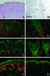
Skin morphology and immunofluorescence study. A: H&E staining. Hyperkeratosis with focal parakeratosis and regular acanthosis with broad rete ridges are observed. Granular layer was lost and vacuolar change of cytoplasm was seen in the upper spinous layer. B: Electron microscopy in the patient’s skin (granular layer of the epidermis). The gap junctions have a typical pentalaminar structure, 20 nm in width. Abnormal junctional structures were not found. D: Desmosome, G: gap junction. C–F: Cx26 and Cx43 expression of the patient’s epidermis. Green (fluorescein isothiocyanate) indicates Cx26 expression. Red (propidium iodide) is nuclear staining. C: Cx26 was expressed in the keratinocytes in the widened rete ridge. D: The staining of the Cx26 in the keratinocytes was more cytoplasmic than membranous, although punctate membranous staining was also seen in the acrosyringium cells. Cx43 expression was observed in the keratinocytes in the middle and upper epidermal layers (E) and was mostly membranous (F). G: Cx26 staining in normal skin. There was no expression of Cx26 in the normal epidermis. The staining at the corneum seemed to be nonspecific. H: Cx43 staining in normal skin. The staining was observed at the membrane of epidermal keratinocytes. Scale bar = 100 nm.
Figure 4-6927
Figure 4-6927
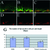
Dye transfer assay of cultured keratinocyte. A: NHEK. B: HaCaT. C: Keratinocytes of KID syndrome. The diffusion of Lucifer yellow of patient’s keratinocytes was less than in NHEK and HaCaT cells. Rhodamine dextran (red) is observed at the edge of the scratched plane because the rhodamine dextran molecule is too bulky to pass through gap junctions (D–F: NHEK, HaCaT, and patient’s keratinocyte, respectively). G: The number of Lucifer yellow-positive cells per unit length of scratched plane. The number of positive cells of the patient’s keratinocytes was significantly smaller than that of NHEK and HaCaT. NHEK: 7.17 ± 1.14 (average ± SD); HaCaT: 7.43 ± 1.07; patient’s cell: 4.34 ± 0.75. *P < 0.01 by Student’s t-test.
Figure 5-6927
Figure 5-6927
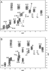
Comparison of the fingerprint region from DQF-COSY spectra of wild type (A) and N14Y mutant (B). The mutation does not cause significant change in chemical shifts when compared to wild type.
Figure 6-6927
Figure 6-6927
Chemical shift differences of amino acid residues in wild type and N14Y mutant. A: The differences in chemical shift defined as δ (wild-type or mutant peptide)-δ (random coil) are plotted. B: The differences in chemical shift defined as wild-type peptide-mutant are plotted.
Figure 7-6927
Figure 7-6927
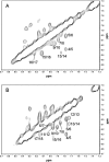
NH-NH regions from NOESY spectra of wild-type (A) and N14Y mutant (B) peptides. Sequential NH-NH cross-peaks are annotated.
Figure 8-6927
Figure 8-6927
Novel NOE signals in N14Y mutant.
Figure 9-6927
Figure 9-6927
Far-UV CD spectra of wild-type and mutant peptides. Wild-type (blue) and mutant peptide (red) were measured in water (A) and in various TFE concentrations (B, C). The numbers indicate the percentage of TFE.
Figure 10-6927
Figure 10-6927
The Cx26 mutations of syndromic sensorineural deafness reported in the literature. The mutations of KID syndrome are aggregated to the N-terminal side of Cx26, especially in the cytoplasmic portion.

References

    1. Kelsell DP, Dunlop J, Hodgins MB. Human diseases: clues to cracking the connexin code? Trends Cell Biol. 2001;11:2–6. - PubMed
    1. Rabionet R, Lopez-Bigas N, Arbones ML, Estivill X. Connexin mutations in hearing loss, dermatological and neurological disorders. Trends Mol Med. 2002;8:205–212. - PubMed
    1. Maestrini E, Korge BP, Ocana-Sierra J, Calzolari E, Cambiaghi S, Scudder PM, Hovnanian A, Monaco AP, Munro CS. A missense mutation in connexin26, D66H, causes mutilating keratoderma with sensorineural deafness (Vohwinkel’s syndrome) in three unrelated families. Hum Mol Genet. 1999;8:1237–1243. - PubMed
    1. Richard G, Rouan F, Willoughby CE, Brown N, Chung P, Ryynanen M, Jabs EW, Bale SJ, DiGiovanna JJ, Uitto J, Russell L. Missense mutations in GJB2 encoding connexin-26 cause the ectodermal dysplasia keratitis-ichthyosis-deafness syndrome. Am J Hum Genet. 2002;70:1341–1348. - PMC - PubMed
    1. Richard G, White TW, Smith LE, Bailey RA, Compton JG, Paul DL, Bale SJ. Functional defects of Cx26 resulting from a heterozygous missense mutation in a family with dominant deaf-mutism and palmoplantar keratoderma. Hum Genet. 1998;103:393–399. - PubMed

Publication types

MeSH terms