. 2006 Aug;169(2):459-70.
doi: 10.2353/ajpath.2006.050969.

Mechanotransduction of extracellular signal-regulated kinases 1 and 2 mitogen-activated protein kinase activity in smooth muscle is dependent on the extracellular matrix and regulated by matrix metalloproteinases

Affiliations

Mechanotransduction of extracellular signal-regulated kinases 1 and 2 mitogen-activated protein kinase activity in smooth muscle is dependent on the extracellular matrix and regulated by matrix metalloproteinases

Karen J Aitken et al. Am J Pathol. 2006 Aug.

Abstract

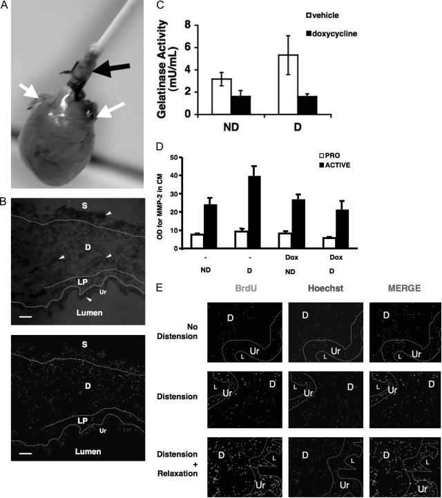
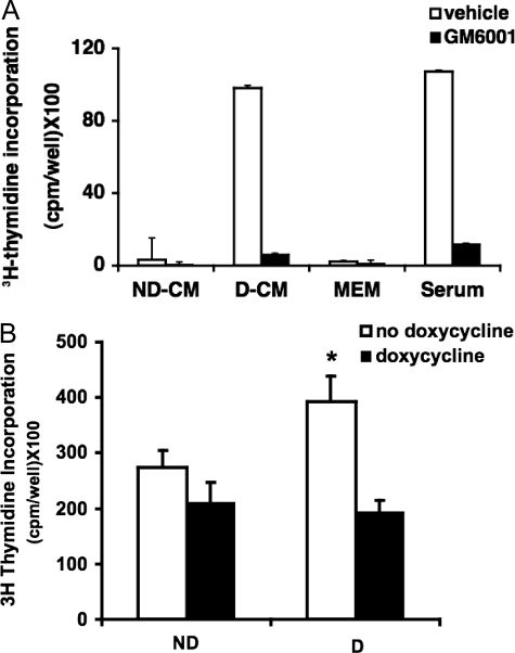
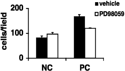
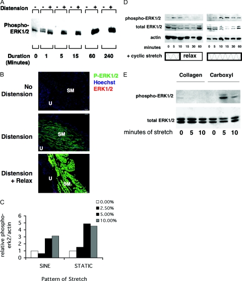
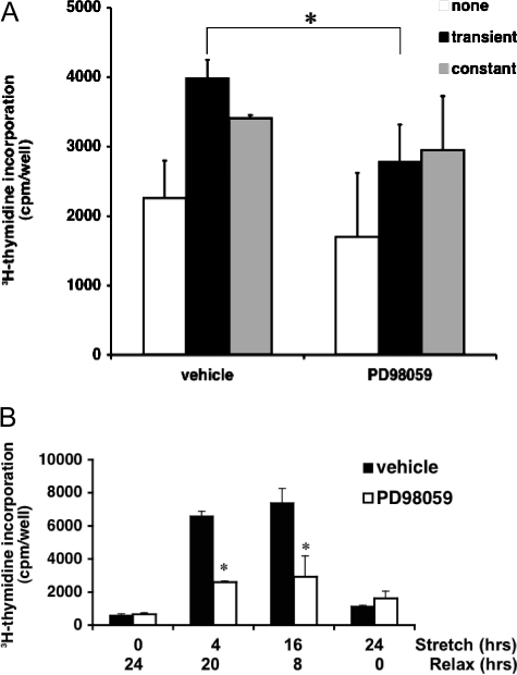
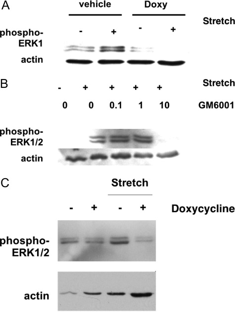
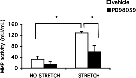
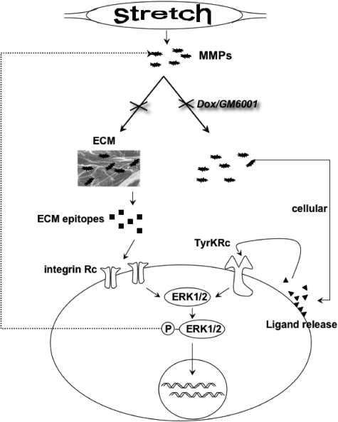
Excessive wall stretch of distensible hollow organs in cardiovascular and urinary systems can activate matrix metalloproteinases (MMPs), thereby releasing matrix neoepitopes and growth factor ligands, leading to ERK1/2 activation. However, the role of MMPs in mechanotransduction of ERK1/2 signaling in the bladder is unknown. We examined bladders undergoing sustained distension over time, which provides a novel platform for smooth muscle mechanotransduction studies. Bladder distension ex vivo caused increased proliferation and MMP activity. Conditioned medium from distended compared with undistended bladders induced proliferation in bladder smooth muscle cells (BSMCs). When conditioned medium from distended bladders was used to proteolyze collagen type I matrices, matrices augmented BSMC proliferation, which was inhibited if bladders were distended in presence of broad-spectrum MMP inhibitors. Distension of ex vivo bladders also induced ERK1/2 phosphorylation in situ, which was dependent on MMP activity in the intact bladder. Similarly, stretching BSMCs in vitro induced increases in ERK1/2 activation and ERK1/2-dependent proliferation under discrete mechanical conditions, and distension conditioned medium itself induced MMP-dependent ERK1/2 activation in BSMCs. Overall, stretch-induced proliferation and ERK1/2 signaling in bladder tissue and BSMCs likely depend on secreted MMP activity. Identification of intermediaries between MMPs and ERK1/2 may elaborate novel mechanisms underlying mechanotransduction in bladder smooth muscle.


Figures

Figure 1-6940
Figure 1-6940
A:Ex vivo model of bladder distension. Bladders of female Sprague-Dawley rats (100–120 g) were surgically exposed, and ureters were ligated. Bladders were catheterized per urethra and removed with sutures that firmly held the catheters in the urethra. For blocking, 1-hour pre-incubations were performed in medium containing the relevant inhibitors. After blocking, bladders were distended by filling the bladder with culture medium to 40 cm of hydrostatic pressure using water manometry or were not distended. Sham controls included ligated, catheterized, but uninflated bladders. White arrows indicate the ligated ureters. Black arrows point to the catheterized urethra, which has been sutured to hold the catheter in place during distension. B: Bladder hyperdistension stimulates in situ gelatinase activity. Ex vivo bladders were examined by in situ zymography. FITC-gelatin layered onto cryosections of ex vivo bladders is digested by MMP activity. Nonfluorescent dark zones (arrowheads) represent digestion of the FITC-gelatin, localizing regions of gelatinase A/B activity. Bottom panel: Hoechst stain of serial section to show nuclei. Ur, urothelium; LP, lamina propria; D, detrusor; S, serosa. Original magnification, ×125. C: Net gelatinase activity of bladders is increased during distension ex vivo. Gelatinases in the conditioned medium released the quenched activity of FITC in the DQ-gelatin. Doxycycline inhibited the increase in gelatinase A/B activity from distended bladders, P = 0.0126; n = 3. D: Secretion of active MMP-2 is increased in distended bladders and inhibited by doxycycline. Pro- and active-MMP-2 are analyzed by Western blotting in the conditioned medium of distended (D) and nondistended (ND) bladders +/− Doxycycline (Dox) pretreatment for 1 hour. E: Bladder distension stimulates in situ BSMC proliferation. BSMC proliferation was assessed by BrdU incorporation and detected using anti-BrdU and anti-mouse-Alexa 488 antibody on cryosections. Bladders were distended by hydrodistension in serum-free medium with BrdU or were not distended. Distended bladders showed increased BrdU incorporation compared with undistended. Ur, uroepithelium; D, detrusor smooth muscle layer; L, lumen. n = 3. Original magnification, ×125.
Figure 2-6940
Figure 2-6940
Bioactivity of conditioned medium (CM) from ex vivo distended bladders is dependent on MMP activity. A: BSMC proliferation is induced by CM-remodeled matrix and is blocked when MMPs are inhibited with GM6001. CM from bladders distended (D-CM) or not (ND-CM) for 24 hours were allowed to proteolyze collagen type I gels in the presence or absence of GM6001. Proliferation was assayed by thymidine incorporation of BSMCs incubated on proteolyzed matrices for 24 hours. FCS and MEM were used as positive and negative controls, respectively; n = 3. B: Bladders were pre-incubated in the MMP inhibitor doxycycline, or vehicle, for 1 hour and then distended for 15 minutes or undistended. BSMCs were incubated in bladder CM plus [3H]thymidine for 72 hours to assess proliferative responses. Inhibition of MMP activity before bladder distension abolished increased proliferative responses of BSMCs; n = 3. *P < 0.05.
Figure 3-6940
Figure 3-6940
Proliferation of BSMCs on proteolyzed matrix is ERK1/2 dependent. Native type I collagen gels (NC) were treated with MEM (NC) or proteolyzed with FCS (PC) overnight and then washed extensively in MEM. BSMCs (n = 3) were plated on matrices. After 24 hours of culture, trypan blue-excluding cells were counted. Proliferation was induced by FCS-proteolyzed gels and was blocked by addition of PD98059.
Figure 4-6940
Figure 4-6940
Distension activates ERK1/2 in whole intact bladder as well as BSMCs. A: ERK1/2 activation in whole distended bladder over a time course from 1 to 240 minutes. Western blotting of bladder tissue protein lysates with phospho-ERK1/2 antibody shows increased P-ERK1/2 after distension; n = 3. B: Localization of ERK1/2 phosphorylation was determined by immunostaining using anti-phospho-ERK1/2 and anti-mouse-Alexa-488 (green). Total ERK1/2 was localized using anti-ERK1/2 and anti-rabbit-Cy3 (red). Nuclei were counterstained with Hoechst (blue). Bladders were cultured ex vivo for 24 hours including 0 minutes (n = 3), 10 minutes, or 24 hours of distension (n = 3). The 10-minute distended bladders were relaxed (deflated) for the rest of the 24-hour period (stretch + relax). Phospho-ERK1/2 appears most strongly in the distension plus relax group in the smooth muscle compartment (SM). Total ERK1/2 can be seen best in the urothelium (U) in these merged images, although it appears throughout when seen on the red channel only. Original magnification, ×125. CE: Stretch parameters alter ERK1/2 activation in vitro. After adherence, BSMCs were serum starved and mechanically stretched on Flexcell plates under various conditions, and their protein was analyzed by Western blotting for phospho- and total ERK1/2 and actin. C: BSMCs were stretched using a cyclic sine pattern or a static pattern at indicated percentages of elongation (n = 3). D: Cells were stretched using a cyclic stretch program (5 seconds on, 5 seconds off) with a sinusoidal pattern on carboxyl-coated plates either transiently (5-minute cyclic stretch + 55-minute relaxation) or continuously (60-minute cyclic stretch) on carboxyl plates (n = 2). E: Cells were stretched on collagen or carboxyl plates with a cyclic stretch pattern as above (n = 3).
Figure 5-6940
Figure 5-6940
Stretch plus a relaxation period is favored by ERK1/2-mediated BSMC proliferation. BSMCs were stretched and then relaxed over defined short- and long-term time periods, in the presence and absence of the MEK inhibitor PD98059. Proliferation was assayed by tritiated thymidine incorporation. A: Over 4 hours (versus unstretched controls), proliferation was preferentially stimulated (up to ∼75%; white versus black bars; P < 0.05) by stretch + relaxation. Constant stretch produced less proliferation (∼40%; white versus gray bars) (n = 3). B: Over 24 hours (versus unstretched controls), proliferation was enhanced nearly 800% during stretch + relaxation (P < 0.05) versus 24-hour continuous stretch (up to ∼50%) or no stretch (0-hour stretch) controls. Proliferation stimulated by a stretch + relaxation pattern was significantly inhibited by blocking ERK1/2 with PD98059 (*P < 0.05 in A; *P < 0.005 in B). PD98059 did not reduce proliferation in continuously stretched cells; n = 3.
Figure 6-6940
Figure 6-6940
MMPs mediate stretch-induced ERK1/2 activation. A: ERK1/2 activation is seen in response to stretch injury and is blocked by addition of doxycycline. Bladders (n = 3) were stretched 15 minutes ex vivo after a 1-hour incubation in doxycycline (doxy) or vehicle control (EtOH). B: Similarly, GM6001 treatment decreases ERK1/2 activation in response to stretch injury. Bladders (n = 3) were stretched for 15 minutes ex vivo after a 1-hour incubation in increasing concentrations of GM6001 or vehicle control (NS, 0). C: Conditioned medium from distended bladders also induced MMP-dependent ERK1/2 phosphorylation in BSMCs. MMP inhibition by doxycycline prevented ERK1/2 activation in BSMC response to distension-conditioned medium. Bladders (n = 3) were distended ex vivo in the presence of doxycycline or were undistended. Conditioned medium was added in vitro to BSMCs for 5 minutes, and BSMC protein was analyzed by Western blotting.
Figure 7-6940
Figure 7-6940
Stretch-induced gelatinase activity is dependent on ERK1/2 activity. Net MMP activity was assayed using the Enzcheck gelatinase activity kit (Molecular Probes). MMP activity in stretched BSMCs was decreased when ERK1/2 was blocked by an MEK inhibitor. BSMCs (n = 3) were stretched with and without PD98059 (25 μmol/L) for 24 hours or were unstretched, and conditioned medium was collected. Gelatinases in the conditioned medium released the quenched activity of FITC in DQ-gelatin, which was compared with a standard curve (*P < 0.01).
Figure 8-6940
Figure 8-6940
Schematic of stretch-induced ERK1/2 activation in BSMCs. Both extracellular and cellular mechanisms of mechanotransduction involving MMPs are induced during stretch injury, which may involve integrins and tyrosine kinase receptors, respectively.

Similar articles

Cited by

References

    1. Capolicchio G, Aitken KJ, Gu JX, Reddy P, Bagli DJ. Extracellular matrix gene responses in a novel ex vivo model of bladder stretch injury. J Urol. 2001;165:2235–2240. - PubMed
    1. Levin R, Levin S, Zhao Y, Buttyan R. Cellular and molecular aspects of bladder hypertrophy. Eur Urol. 1997;32(Suppl 1):15–21. - PubMed
    1. Polyakova V, Hein S, Kostin S, Ziegelhoeffer T, Schaper J. Matrix metalloproteinases and their tissue inhibitors in pressure-overloaded human myocardium during heart failure progression. J Am Coll Cardiol. 2004;44:1609–1618. - PubMed
    1. Iwanaga Y, Aoyama T, Kihara Y, Onozawa Y, Yoneda T, Sasayama S. Excessive activation of matrix metalloproteinases coincides with left ventricular remodeling during transition from hypertrophy to heart failure in hypertensive rats. J Am Coll Cardiol. 2002;39:1384–1391. - PubMed
    1. Sutherland RS, Baskin LS, Elfman F, Hayward SW, Cunha GR. The role of type IV collagenases in rat bladder development and obstruction. Pediatr Res. 1997;41:430–434. - PubMed

Publication types

MeSH terms

LinkOut - more resources