

Elevated cysteine-rich 61 mediates aberrant collagen homeostasis in chronologically aged and photoaged human skin
- PMID: 16877350
- PMCID: PMC1698795
- DOI: 10.2353/ajpath.2006.060128
Elevated cysteine-rich 61 mediates aberrant collagen homeostasis in chronologically aged and photoaged human skin
Abstract
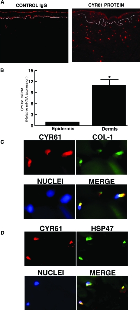
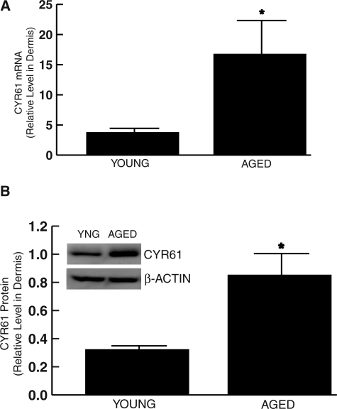
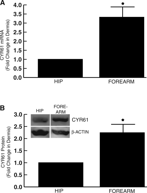
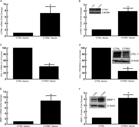
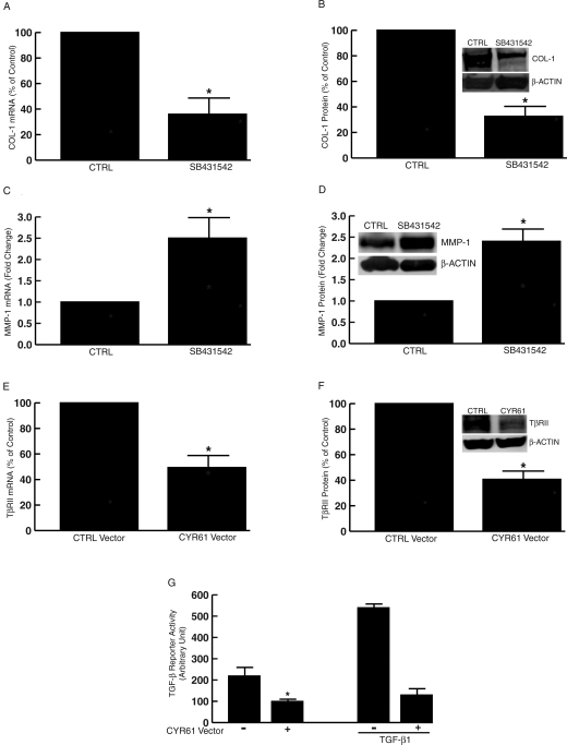
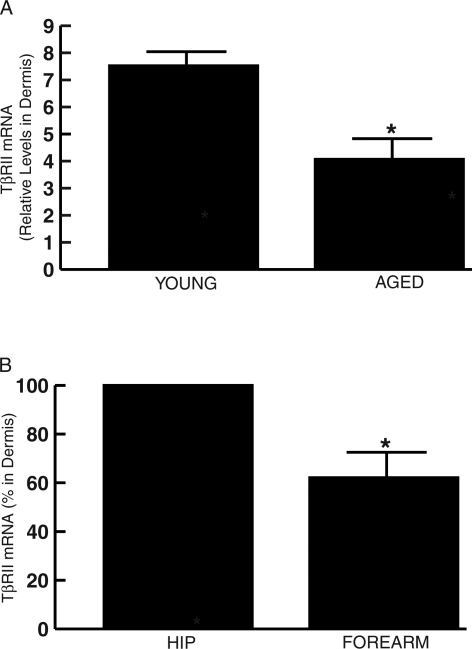
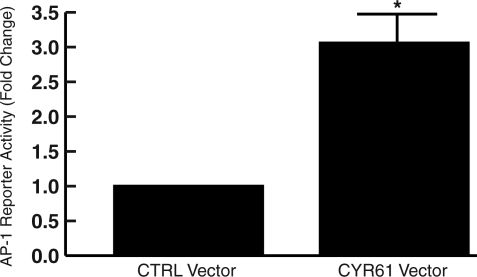
Alterations of human skin connective tissue structure and function are prominent features of chronological aging and solar UV irradiation-induced premature aging (photoaging). These skin connective tissue abnormalities result, in part, from reduced synthesis and elevated degradation of type I collagen, the major structural protein in skin. Here, we report that cysteine-rich 61 (CYR61/CCN1), a novel mediator of collagen homeostasis, is predominantly expressed in human skin connective tissue and is significantly elevated in fibroblasts in chronologically aged (80+ years) and photoaged human skin in vivo. In cultured human skin fibroblasts, elevated CYR61 expression substantially reduces type I procollagen and concurrently increases matrix metalloproteinase-1 (MMP-1), which initiates fibrillar collagen degradation. Elevated CYR61 caused down-regulation of transforming growth factor-beta type II receptor mRNA and protein levels, thereby impairing the transforming growth factor-beta pathway, which reduced type I procollagen and raised MMP-1 expression. Furthermore, elevated CYR61 induced transcription factor activator protein-1 (AP-1), which functions to stimulate MMP-1 expression. Thus, elevated expression of CYR61 in human skin fibroblasts acts through multiple pathways to cause alterations of collagen homeostasis similar to those pathways observed in aged human skin in vivo. These data identify CYR61 as a pivotal regulator of collagen production and degradation in aged and photoaged human skin.
Figures
Similar articles
-
Retinoids suppress cysteine-rich protein 61 (CCN1), a negative regulator of collagen homeostasis, in skin equivalent cultures and aged human skin in vivo.Exp Dermatol. 2011 Jul;20(7):572-6. doi: 10.1111/j.1600-0625.2011.01278.x. Epub 2011 Apr 13. Exp Dermatol. 2011. PMID: 21488975 Free PMC article.
-
Ultraviolet irradiation induces CYR61/CCN1, a mediator of collagen homeostasis, through activation of transcription factor AP-1 in human skin fibroblasts.J Invest Dermatol. 2010 Jun;130(6):1697-706. doi: 10.1038/jid.2010.29. Epub 2010 Feb 18. J Invest Dermatol. 2010. PMID: 20164845
-
Solar ultraviolet irradiation reduces collagen in photoaged human skin by blocking transforming growth factor-beta type II receptor/Smad signaling.Am J Pathol. 2004 Sep;165(3):741-51. doi: 10.1016/s0002-9440(10)63337-8. Am J Pathol. 2004. PMID: 15331399 Free PMC article.
-
Mechanical regulation of the Cyr61/CCN1 and CTGF/CCN2 proteins.FEBS J. 2006 Aug;273(16):3639-49. doi: 10.1111/j.1742-4658.2006.05360.x. Epub 2006 Jul 19. FEBS J. 2006. PMID: 16856934 Review.
-
UV-light-induced signal cascades and skin aging.Ageing Res Rev. 2002 Sep;1(4):705-20. doi: 10.1016/s1568-1637(02)00024-7. Ageing Res Rev. 2002. PMID: 12208239 Review.
Cited by
-
Enhancing structural support of the dermal microenvironment activates fibroblasts, endothelial cells, and keratinocytes in aged human skin in vivo.J Invest Dermatol. 2013 Mar;133(3):658-667. doi: 10.1038/jid.2012.364. Epub 2012 Oct 25. J Invest Dermatol. 2013. PMID: 23096713 Free PMC article.
-
How much do we really know about our favorite cosmeceutical ingredients?J Clin Aesthet Dermatol. 2010 Feb;3(2):22-41. J Clin Aesthet Dermatol. 2010. PMID: 20725560 Free PMC article.
-
Structural and Functional Changes and Possible Molecular Mechanisms in Aged Skin.Int J Mol Sci. 2021 Nov 19;22(22):12489. doi: 10.3390/ijms222212489. Int J Mol Sci. 2021. PMID: 34830368 Free PMC article. Review.
-
The matricellular protein CCN1 induces fibroblast senescence and restricts fibrosis in cutaneous wound healing.Nat Cell Biol. 2010 Jul;12(7):676-85. doi: 10.1038/ncb2070. Epub 2010 Jun 6. Nat Cell Biol. 2010. PMID: 20526329 Free PMC article.
-
Reduction of fibroblast size/mechanical force down-regulates TGF-β type II receptor: implications for human skin aging.Aging Cell. 2016 Feb;15(1):67-76. doi: 10.1111/acel.12410. Epub 2015 Oct 8. Aging Cell. 2016. PMID: 26780887 Free PMC article.
References
-
- Lau L, Lam SC-T. The CCN family of angiogenic regulators: the integrin connection. Exp Cell Res. 1999;248:44–57. - PubMed
-
- Brigstock D. The connective tissue growth factor/cysteine-rich 61/nephroblastoma overexpressed (CCN) family. Endocr Rev. 1999;20:189–206. - PubMed
-
- Chen C-C, Mo F-E, Lau L. The angiogenic factor Cyr61 activates a genetic program for wound healing in human skin fibroblasts. J Biol Chem. 2001;276:47329–47337. - PubMed
Publication types
MeSH terms
Substances
Grants and funding
LinkOut - more resources
Full Text Sources
Medical
Research Materials
