. 2006 Aug;169(2):503-14.
doi: 10.2353/ajpath.2006.050960.

p62 ubiquitin binding-associated domain mediated the receptor activator of nuclear factor-kappaB ligand-induced osteoclast formation: a new insight into the pathogenesis of Paget's disease of bone

Affiliations

p62 ubiquitin binding-associated domain mediated the receptor activator of nuclear factor-kappaB ligand-induced osteoclast formation: a new insight into the pathogenesis of Paget's disease of bone

Kirk H M Yip et al. Am J Pathol. 2006 Aug.

Abstract

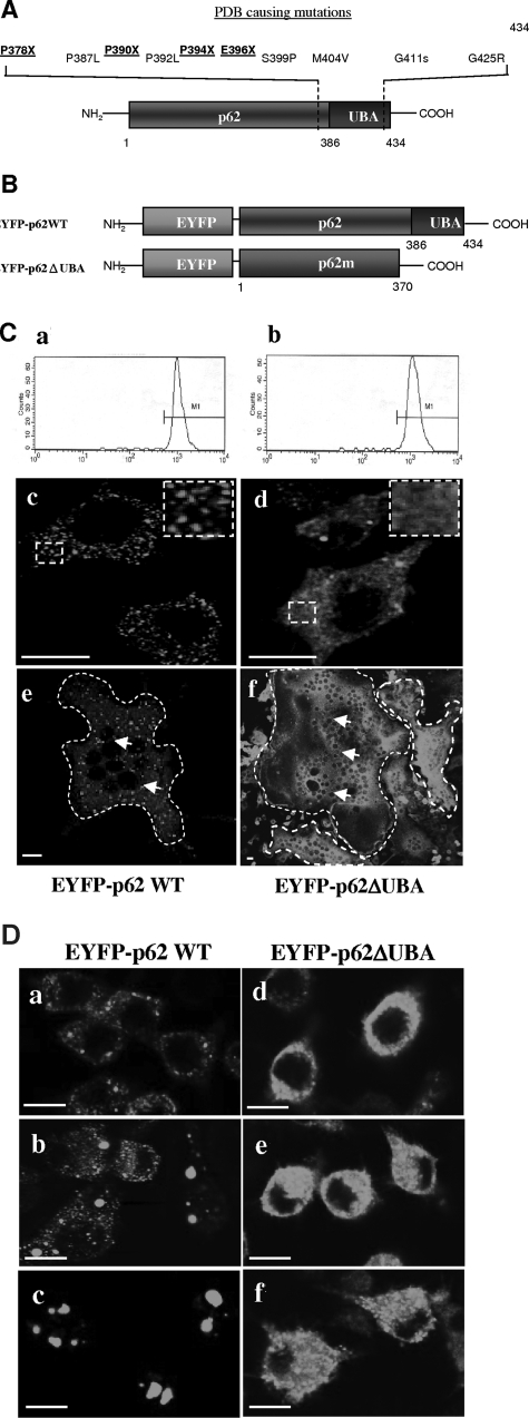
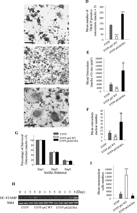
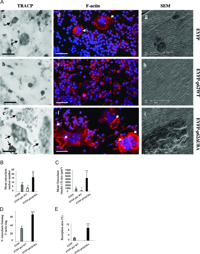
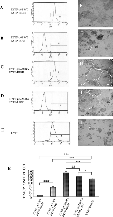
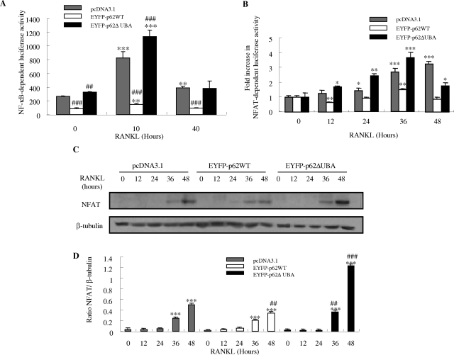
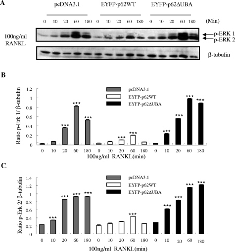
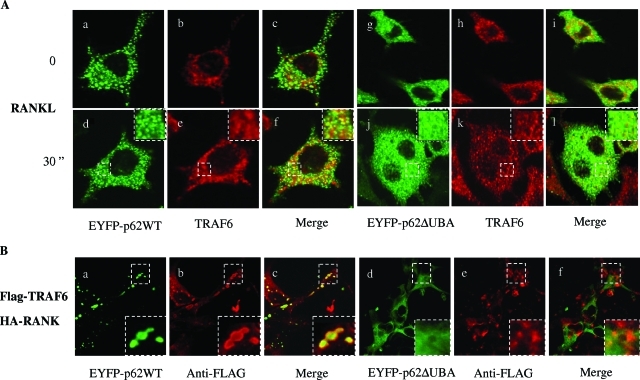
Paget's disease of bone (PDB) is a debilitating bone disorder characterized by giant osteoclasts, enhanced bone destruction, and irregular bone formation. Recently, mutations in SQSTM1 (also known as p62) have been detected in PDB sufferers, with all mutations resulting in either loss of function or truncation/deletion of the ubiquitin binding-associated (UBA) domain. We hypothesized that mutation in the p62 gene resulting in either deletion or premature termination of the UBA domain accounts for the elevated osteoclastic formation and bone resorption associated with PDB. Remarkably, overexpression of the p62 UBA domain deletion mutant (p62DeltaUBA) significantly enhanced osteoclastogenesis in vitro compared to cells expressing either wild-type p62 (p62WT) or a control vector in a RAW264.7 osteoclastogenic system. Overexpression of p62DeltaUBA potentiated the formation of abnormally large multinucleated osteoclasts and resorption of bone, reminiscent of PDB. Consistent with the enhancement of osteoclastogenesis, overexpression of p62DeltaUBA potentiated receptor activator of nuclear factor-kappaB ligand-induced activation of nuclear factor-kappaB, NFAT, and ERK phosphorylation. Furthermore, as determined by confocal microscopy, deletion of the p62 UBA domain impaired the association of p62 with TRAF6 in the proteasomal compartment. These results suggest that the UBA domain encodes essential regulatory elements required for receptor activator of nuclear factor-kappaB ligand-induced osteoclast formation and bone resorption that may be directly associated with the progression of PDB.


Figures

Figure 1-6923
Figure 1-6923
Expression and subcellular localization of p62 and the UBA domain deletion mutant. A: Schematic representation of the detected PDB-causing p62 UBA domain mutations. Four of the PDB mutations (K378X, A390X, L394X, and E396X) lead to introduction of stop codons that result in premature termination of the translation of p62 UBA domain.B: Schematic illustration of EYFP-tagged p62 wild-type (EFYP-p62WT) and p62 UBAdomain deletion mutant (EYFP-p62ΔUBA) constructs. C: FACS analysis of postsorted RAW264.7 cells stably expressing EYFP-p62WT (a) or EYFP-p62ΔUBA (b). Confocal images of the cellular localization of EYFP-p62WT (c) or EYFP-p62ΔUBA (d) in RAW264.7 stable cell lines cultured on glass coverslips. Confocal images of the cellular localization of EYFP-p62WT (e) or EYFP-p62ΔUBA (f) in OCLs. Borders of OCLs are outlined with dashed lines and nuclei are marked with arrows. In brief, RAW264.7 cell lines cultured on glass coverslips were treated with 100 ng/ml of RANKL for 5 days and fixed. EYFP signals in EYFP-p62WT- and EYFP-p62ΔUBA-expressing OCLs were analyzed by confocal microscopy. D: The requirement of p62 UBA domain for subcellular distribution of p62 to the proteasomal degradation pathway. EYFP-p62WT and EYFP-p62ΔUBA in stable RAW264.7 cell lines were grown on glass coverslips and either left untreated or treated with MG132 as indicated. The cells were fixed with 4% paraformaldehyde and processed for confocal microscopy. Representative confocal images of the cellular localization are shown. a–c: Localization of EYFP-p62WT in the absence of MG132 (a), treated with 0.5 μm MG132 for 24 hours (b), or treated with 2 μm MG132 for 12 hours (c). d–f: Localization of EYFP-p62ΔUBA in the absence of MG132 (d), treated with 0.5 μm MG132 for 24 hours (e), or treated with 2 μm MG132 for 12 hours (f). Insets represent magnifications of hatched regions. Scale bars = 10 μm.
Figure 2-6923
Figure 2-6923
The effect of overexpression of EYFP-p62WT or EYFP-p62ΔUBA on osteoclast size, multinucleation, and survival. RAW264.7 cells stably expressing EYFP-p62WT or EYFP-p62ΔUBA were cultured in the presence of RANKL. After 5 days, the cells were fixed with 4% paraformaldehyde and stained for TRACP activity. A–C: Representative images of TRACP staining of OCLs from one of the three experiments are shown. D–F: Quantitative analysis shows the mean number of TRACP-positive OCLs (D), mean OCLs sizes (E), and mean OCLs nuclei numbers (F). G: Similar experiments were performed in which RAW264.7 cells stably expressing EYFP-p62WT, EYFP-p62ΔUBA, or EYFP were cultured in the presence of 100 ng/ml RANKL for 5 days. Fresh medium with no RANKL was then replaced and cells were continued in culture for an additional 1, 3, and 5 days. Cells were fixed, and the number of surviving osteoclasts (nuclei >3) were counted. The effect of overexpression of p62WT, EYFP-p62ΔUBA, or EYFP fusion proteins on OCL survival was quantified by the ratio between the OCLs remained on the third or fifth day after RANKL withdrawal and the initial number of osteoclasts present on the first day of RANKL withdrawal. H: Representative results from three independent experiments showing semiquantitative RT-PCR analysis of the effect of overexpression of p62WT or p62ΔUBA on DC-STAMP gene expression during RANKL-induced RAW cell differentiation. I: The total number of mononuclear cells presented in p62WT, EYFP-p62ΔUBA, and EYFP cultures after 5 days of RANKL stimulation were counted. Four representative views of images were taken from cultures. *P < 0.05; **P < 0.01; ***P < 0.001. Scale bars = 50 μm.
Figure 3-6923
Figure 3-6923
The effect of overexpression of EYFP-p62WT or EYFP-p62ΔUBA on osteoclast resorptive activity. EYFP-p62WT, EYFP-p62ΔUBA, or EYFP stably expressing RAW264.7 cell lines were cultured on bovine bone slices in the presence of RANKL (100 ng/ml). A: After 14 days, cells were fixed and stained for either TRACP (a–c) or rhodamine-phalloidin (F-actin) (d–f) and visualized by light microscopy and confocal microscopy, respectively. Cells were then removed, and resorptive lacunae were assessed by scanning electron microscopy (g–i). F-Actin rings are marked with arrows. Quantitative analysis shows the mean nuclei numbers (B) and sizes (C) of osteoclasts, percentage of osteoclasts forming F-actin rings on bovine bone slice (D), and percentage of bovine bone slice surface occupied by resorption lacunae (E). *P < 0.05; **P < 0.01; ***P < 0.001. Scale bars = 50 μm.
Figure 4-6923
Figure 4-6923
The levels of overexpression of EYFP-p62WT or EYFP-p62ΔUBA affect osteoclastogenesis. A–E: FACS analyses of EYFP-p62WT, EYFP-p62ΔUBA, and EYFP control. RAW264.7 cells stably expressing EYFP-p62WT or EYFP-p62ΔUBA were FACS-sorted into two populations, EYFP-HIGH and EYFP-LOW. Stable RAW264.7 were treated with RANKL for 5 days, fixed, and subjected to TRACP staining. F–J: Representative images of TRACP staining from one of the three experiments are shown. K: Quantitative analysis was preformed by counting the total number of OCLs. P value indicates the difference between respective controls, *P < 0.05, ##P < 0.001, ***/###P < 0.001.
Figure 5-6923
Figure 5-6923
The effects of overexpression of EYFP-p62WT or EYFP-p62ΔUBA on RANKL-induced NF-κB and NFAT-dependent transcription. RAW264.7 cells stably expressing EYFP-p62WT or EYFP-p62ΔUBA were transfected with the 3-kb Luc-SV40 NF-κB luciferase reporter plasmid (A) or pNFAT-TA Luc reporter gene (B) and treated with 100 ng/ml of RANKL for various time periods. After stimulation, cells were lysed and luciferase activities in lysates determined. Each bar is the mean ± SEM from triplicate wells. Representative results from three independent experiments are shown. *The P values of the effect of RANKL stimulation compared to its respective controls (***P < 0.001, **P < 0.01, *P < 0.05). #The P values of the effect of overexpression of pEYFP-62WT and pEYFP-62ΔUBA on RANKL-induced NF-κB activity compared to pcDNA3.1-transfected control (###P < 0.001). C–D: Effect of EYFP-p62WT and EFYP-p62ΔUBA overexpression on RANKL-induced NFAT protein expression in RAW stable cell lines. RAW264.7 cells stably transfected with pcDNA3.1, EYFP-p62WT, and EFYP-p62ΔUBA were stimulated with 100 ng/ml of RANKL for the indicated times. After stimulation, whole cell extracts were analyzed for NFAT protein expression by Western blotting. The same membrane was stripped and reprobed with an antibody for β-tubulin, which served as an internal control for differences in loading and transfer. C: The levels of NFAT and β-tubulin proteins are shown. D: NFAT protein levels are shown as the ratio of NFAT to β-tubulin. Each bar is the mean ± SEM from representative results from three independent experiments. *The P values of the effect on RANKL-induced NFAT protein synthesis at different time points compared to its unstimulated control (***P < 0.001). #The P value compared to the pcDNA3.1 control (##P < 0.01, ###P < 0.001).
Figure 6-6923
Figure 6-6923
The effects of EYFP-p62WT and EFYP-p62ΔUBA overexpression on RANKL-induced phosphorylation of ERK in RAW stable cell lines. RAW264.7 cells stably transfected with pcDNA3.1, EYFP-p62WT, and EFYP-p62ΔUBA were stimulated with 100 ng/ml of RANKL for the indicated time. On stimulation, whole cell extracts were analyzed for phosphorylation of ERK 1 and 2 proteins by Western blotting with monoclonal anti-p-ERK antibody at 1/1000 dilution, followed by enhanced chemiluminescence. The same membrane was stripped and reprobed with an antibody for β-tubulin, which served as an internal control for differences in loading and transfer. A: The levels of ERK 1/2, and β-tubulin proteins are shown. B and C: Levels of phosphorylation of ERK proteins are shown as the ratio of phosphorylated ERK to β-tubulin. Each bar is the mean ± SEM from three independent experiments (***P < 0.001).
Figure 7-6923
Figure 7-6923
Deletion of UBA domain disrupts RANKL p62 association with TRAF6. A: RAW264.7 cells stably expressing EYFP-p62WT or EYFP-p62ΔUBA were grown on glass coverslips and either left untreated or treated with RANKL for 30 minutes. The cells were fixed with 4% paraformaldehyde, immunostained with anti-TRAF6 antibody, and processed for confocal microscopy. Representative confocal images of the cellular localization are shown. Co-localization of EYFP-p62WT with TRAF6 in untreated (a–c) or RANKL-treated RAW cells (d–f). Co-localization of EYFP-p62ΔUBA with TRAF6 in untreated (g–i) or RANKL-treated RAW264.7 cells (j–l). B: COS-7 cells were co-transfected with EYFP-p62WT (a–c) or EYFP-p62ΔUBA (d–f), pFLAG-TRAF6, and pHA-RANK for 24 hours. The cells were fixed and immunostained with anti-FLAG antibody, followed by secondary antibody (Alexa Fluor 546 goat anti-mouse IgG). The images were recorded under confocal microscope. Insets represent magnifications of hatched regions.

Similar articles

Cited by

References

    1. Roodman GD. Mechanisms of abnormal bone turnover in Paget’s disease. Bone. 1999;24:39S–40S. - PubMed
    1. Reddy SV. Etiology of Paget’s disease and osteoclast abnormalities. J Cell Biochem. 2004;93:688–696. - PubMed
    1. Layfield R, Hocking LJ. SQSTM1 and Paget’s disease of bone. Calcif Tissue Int. 2004;75:347–357. - PubMed
    1. Morgan SL, Ward L, Ward BK, Walsh J, Xu J: A novel mutation (K378X) in the sequestosome1 gene associated with a sever phenotype in Paget’s disease of bone. International Symposium on Paget’s Disease, 2005 July 8–9, St. Catherine’s College, Oxford, UK - PubMed
    1. Hocking LJ, Lucas GJ, Daroszewska A, Cundy T, Nicholson GC, Donath J, Walsh JP, Finlayson C, Cavey JR, Ciani B, Sheppard PW, Searle MS, Layfield R, Ralston SH. Novel UBA domain mutations of SQSTM1 in Paget’s disease of bone: genotype phenotype correlation, functional analysis, and structural consequences. J Bone Miner Res. 2004;19:1122–1127. - PubMed

MeSH terms