. 2006 Aug;169(2):553-65.
doi: 10.2353/ajpath.2006.051067.

NUB1 suppresses the formation of Lewy body-like inclusions by proteasomal degradation of synphilin-1

Affiliations

NUB1 suppresses the formation of Lewy body-like inclusions by proteasomal degradation of synphilin-1

Kunikazu Tanji et al. Am J Pathol. 2006 Aug.

Abstract

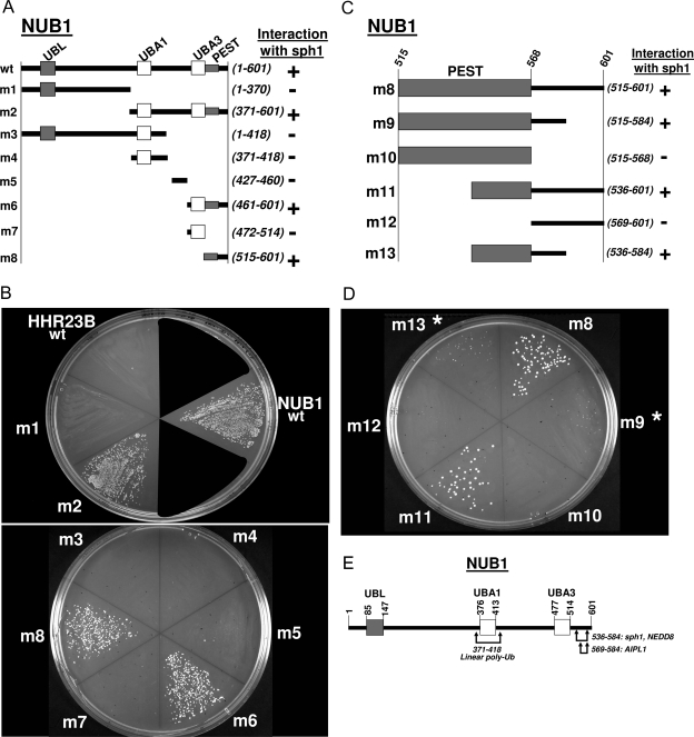
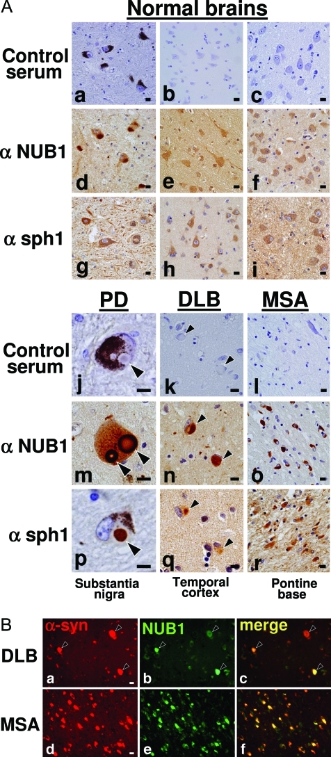
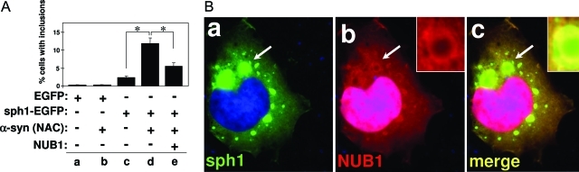
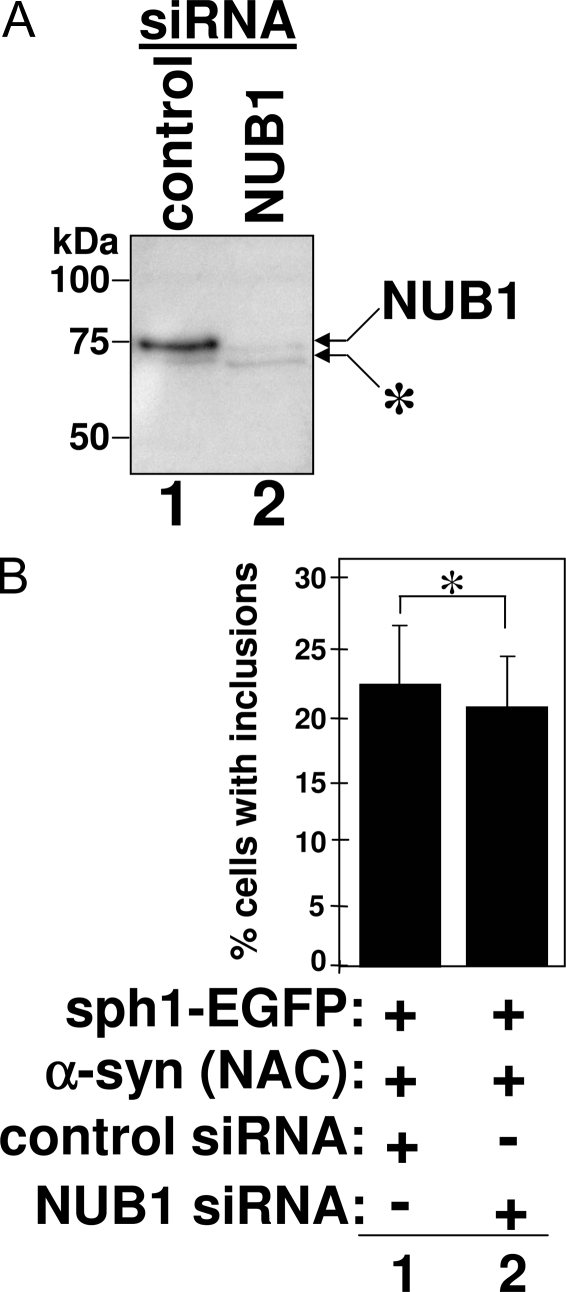
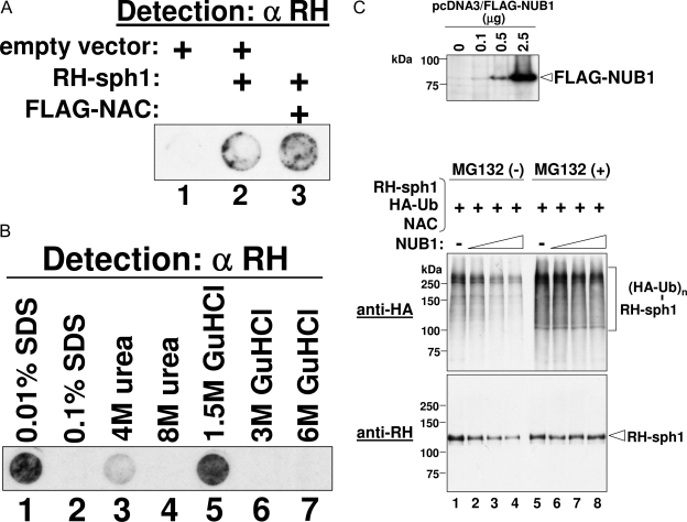
NUB1 is a potent down-regulator of the ubiquitin-like protein NEDD8, because it targets NEDD8 to the proteasome for proteolytic degradation. From results in this study, we found that NUB1 physically interacts with synphilin-1 through its NEDD8-binding site, implying that NUB1 also targets synphilin-1 to the proteasome for degradation. Synphilin-1 is a major component of inclusion bodies found in the brains of patients with neurodegenerative alpha-synucleinopathies, including Parkinson's disease. In this study, we immunostained sections of brains from patients with Parkinson's disease and other alpha-synucleinopathies and demonstrated that NUB1, as well as synphilin-1, accumulates in the inclusion bodies. To define the role of NUB1 in the formation of these inclusion bodies, we performed a co-transfection assay using cultured HEK293 cells. This assay showed that NUB1 suppresses the formation of synphilin-1-positive inclusions. Further, biochemical assays revealed that NUB1 overexpression leads to the proteasomal degradation of synphilin-1. These results and our previous observations suggest that NUB1 indeed targets synphilin-1 to the proteasome for its efficient degradation, which, because of the resultant reduction in synphilin-1, suppresses the formation of synphilin-1-positive inclusions.


Figures

Figure 1-6915
Figure 1-6915
In vitro interaction between NUB1 and synphilin-1. GST and GST-fused HHR23B and NUB1 were expressed in bacteria and purified using glutathione-Sepharose beads. RH-tagged synphilin-1 (sph1) expressed in bacteria was then precipitated with these beads, which were coated with GST alone (lane 2), GST-HHR23B (lane 3), or GST-NUB1 (lane 4). The precipitates were analyzed by Western blotting using anti-RH antibody to detect RH-synphilin-1 (top) and anti-GST antibody to detect immobilized GST, GST-HHR23B, or GST-NUB1 (bottom). In lane 1, the input of RH-synphilin-1 diluted to 1:40 was loaded as a control. Molecular size markers are shown in kilodaltons.
Figure 2-6915
Figure 2-6915
Mapping for the synphilin-1-binding site on NUB1 using a yeast two-hybrid system. A: Summary of interaction between synphilin-1 and truncated NUB1 (m1 to m8). B: Primary data of interaction between synphilin-1 and truncated NUB1 (m1 to m8). The yeast strain AH109 was transformed with pGADT7/synphilin-1 and the pGBKT7 construct encoding wild-type NUB1 (wt), truncated NUB1 (m1 to m8), or HHR23B as a negative control. Transformed yeast cells were grown on a selection plate to determine the specific protein-protein interaction. C: Summary of interaction between synphilin-1 and C-terminal mutant NUB1 (m8 to m13). D: Primary data of interaction between synphilin-1 and C-terminal mutant NUB1 (m8 to m13). The yeast strain AH109 was transformed with pGADT7/synphilin-1 and the pGBKT7 construct encoding truncated NUB1 (m8 to m13). Transformed yeast cells were grown on a selection plate to determine the specific protein-protein interaction. Asterisks indicate low growth of yeast colonies. E: Location of synphilin-1-binding site on NUB1, indicated by arrows. Binding sites of other proteins, such as NEDD8, linear polyubiquitin, and AIPL1, are also indicated by arrows.
Figure 3-6915
Figure 3-6915
Expression of NUB1 in various human tissues. A: Northern blot analysis. mRNA expression of NUB1 (top) and β-actin (bottom) was examined in a variety of normal human tissues. RNA size markers are shown in kilobases. B: Western blot analysis. Protein expression of NUB1 (top), synphilin-1 (middle), and actin (bottom) was examined using a variety of human tissues purchased from ProSci (Poway, CA). Molecular size markers are shown in kilodaltons.
Figure 4-6915
Figure 4-6915
Localization of NUB1 in inclusions of patient brains. A: Localization of NUB1 and synphilin-1 in inclusion bodies of the brains of patients with neurodegenerative α-synucleinopathies. Immunohistochemical studies were performed on the substantia nigra (a, d, and g), temporal cortex (b, e, and h), and pontine base (c, f, and i) from control subjects and the substantia nigra from PD (j, m, and p), temporal cortex from DLB (k, n, and q), and pontine base from MSA (l, o, and r) using control serum (a–c and j–l), anti-NUB1 antibody (d–f and m–o), and anti-synphilin-1 antibody (g–i and p–r). Anti-NUB1 (d–f) and anti-synphilin-1 antibodies (g–i) weakly immunolabeled the neuronal cytoplasm and processes in the normal brains. LBs in the brains of patients with PD and DLB were immunostained with anti-NUB1 (m and n), as were glial cytoplasmic inclusions (GCIs) in patients with MSA (o). LBs and GCIs were also positive for synphilin-1 (p–r). B: Double-immunofluorescence staining showing co-localization of phosphorylated α-synuclein and NUB1 in cortical LBs in patients with DLB (a–c) and GCIs in patients with MSA (d–f). α-Synuclein appears red (a and d) and NUB1 appears green (b and e). The overlap of α-synuclein with NUB1 appears yellow (c and f). Scale bars = 10 μm.
Figure 5-6915
Figure 5-6915
Localization of NUB1 in inclusions of cultured HEK293 cells. A: Synphilin-1-positive inclusions in HEK293 cells transfected with synphilin-1 and α-synuclein. HEK293 cells were transfected with pNAC-Sph1 to express both FLAG-NAC and synphilin-1-EGFP. After 24 hours, the cells were fixed and immunostained with anti-FLAG antibody. The primary antibody was then labeled with Texas Red-conjugated secondary antibody. The immunostained cells were treated with Hoechst 33258 dye for the nuclear staining and then analyzed under a fluorescence microscope. The localization of synphilin-1-EGFP was shown by the green fluorescence of EGFP (a), and the localization of FLAG-NAC was shown by the red fluorescence of Texas Red (b). Their co-localization was shown by the merging of both fluorescences (c). B: Co-localization of synphilin-1 with endogenous proteins related to LBs in inclusions of HEK293 cells. HEK293 cells were transfected with pNAC-Sph1. After 24 hours, the cells were fixed and immunostained with antibodies to NUB1 (a–c), the Rpt5 subunit of PA700 (d–f), the α-subunits of 20S proteasome (g–i), ubiquitin (Ub; j–l), or NEDD8 (m–o). The primary antibody was then labeled with Texas Red-conjugated secondary antibody. The immunostained cells were treated with Hoechst 33258 dye for the nuclear staining and then analyzed under a fluorescence microscope. The localization of synphilin-1-EGFP was shown by the green fluorescence of EGFP (a, d, g, j, and m), and the localization of other endogenous proteins was shown by the red fluorescence of Texas Red (b, e, h, k, and n). Their co-localization with synphilin-1 was shown by the merging of both fluorescences (c, f, i, l, and o). Scale bar = 10 μm.
Figure 6-6915
Figure 6-6915
NUB1-mediated regulation in the formation of synphilin-1-positive inclusions. A: Effect of NUB1 overexpression on the formation of synphilin-1-positive inclusions. In HEK293 cells transfected with various constructs, cytoplasmic inclusions with green fluorescence were quantified. Cells expressing EGFP (a and b) or synphilin-1-EGFP (c–e) were counted to determine the number of transfected cells. Then, the transfected cells containing cytoplasmic inclusions were counted. The value of percent cells with inclusions was calculated as the ratio of the number of transfected cells containing inclusions to the total number of transfected cells. Each bar represents the mean ± SE (*P < 0.0001). B: Location of overexpressed NUB1 in synphilin-1-positive inclusions. As done in Ae, RH-NUB1 was co-expressed with synphilin-1-EGFP and NAC in HEK293 cells. After 24 hours, the cells were fixed and immunostained with anti-RH antibody (1:1600). The primary antibody was then labeled with Texas Red-conjugated secondary antibody (1:400). The immunostained cells were treated with Hoechst 33258 dye for the nuclear staining and then analyzed under a fluorescence microscope. The location of synphilin-1-EGFP was shown by the green fluorescence of EGFP (a), and the location of RH-NUB1 was shown by the red fluorescence of Texas Red (b). Their co-localization was shown by the merging of both fluorescences (c).
Figure 7-6915
Figure 7-6915
RNAi of NUB1. A: Effect of NUB1 siRNA on the expression of endogenous NUB1. HEK293 cells were transfected with a siRNA of control or NUB1 and a plasmid encoding synphilin-1-EGFP and FLAG-NAC. Twenty-four hours after transfection, cells were lysed. The expression level of endogenous NUB1 was then determined by Western blotting using anti-NUB1 antibody. A nonspecific band is indicated by an asterisk. B: Effect of NUB1 siRNA on the formation of synphilin-1-positive inclusions. HEK293 cells were transfected with a siRNA of control or NUB1 and a plasmid encoding both synphilin-1-EGFP and FLAG-NAC. Twenty-four hours after transfection, cells were fixed, and then the transfected cells containing cytoplasmic inclusions were counted under a fluorescence microscope. Each bar represents the mean ± SE (*P = 0.532, not significant).
Figure 8-6915
Figure 8-6915
Reduction of synphilin-1 by overexpression of NUB1 in HEK293 cells. A: Filter-trap assay of synphilin-1-positive inclusions. The inclusions were generated by overexpressing RH-synphilin-1 with or without NAC in HEK293 cells. The inclusions were then trapped on a 0.22-μm cellulose acetate membrane and detected with anti-RH antibody. B: Solubilization of synphilin-1-positive inclusions in various solutions. The inclusions were generated by overexpressing RH-synphilin-1 with NAC in HEK293 cells. Total cell lysate was prepared from the cells in various solutions and analyzed by filter-trap assay. C: NUB1-mediated proteasomal degradation of synphilin-1. By transfecting plasmids, FLAG-NUB1 was overexpressed at various levels with RH-synphilin-1, HA-ubiquitin, and NAC in HEK293 cells. A portion of the cells were lysed for Western blot analysis using anti-FLAG antibody to detect FLAG-NUB1 (top). The rest of the cells were further cultured in the presence or absence of MG132. The cells were then lysed in 6 mol/L of guanidine HCl, and a total cell lysate was prepared. RH-synphilin-1 was precipitated with TALON beads from the total cell lysate and analyzed by Western blotting using anti-HA antibody to detect ubiquitinated RH-synphilin-1 and anti-RH antibody to detect RH-synphilin-1 (middle and bottom). Note: In all samples, 2.5 μg of plasmid DNA for the expression of RH-synphilin-1, HA-ubiquitin, and NAC were transfected into HEK293 cells on a 6-cm dish. In addition, the cells were co-transfected with a plasmid for the expression of FLAG-NUB1 in amounts of 0 μg (lanes 1 and 5), 0.1 μg (lanes 2 and 6), 0.5 μg (lanes 3 and 7), or 2.5 μg (lanes 4 and 8).

Similar articles

Cited by

References

    1. Kamitani T, Kito K, Nguyen HP, Yeh ETH. Characterization of NEDD8, a developmentally down-regulated ubiquitin-like molecule. J Biol Chem. 1997;272:28557–28562. - PubMed
    1. Wada H, Yeh ETH, Kamitani T. Identification of NEDD8-conjugation site in human cullin-2. Biochem Biophys Res Commun. 1999;257:100–105. - PubMed
    1. Stickle NH, Chung J, Klco JM, Hill RP, Kaelin WG, Jr, Ohh M. pVHL modification by NEDD8 is required for fibronectin matrix assembly and suppression of tumor development. Mol Cell Biol. 2004;24:3251–3261. - PMC - PubMed
    1. Xirodimas DP, Saville MK, Bourdon JC, Hay RT, Lane DP. Mdm2-mediated NEDD8 conjugation of p53 inhibits its transcriptional activity. Cell. 2004;118:83–97. - PubMed
    1. Yeh ETH, Gong L, Kamitani T. Ubiquitin-like proteins: new wines in new bottles. Gene. 2000;248:1–14. - PubMed

Publication types

MeSH terms

LinkOut - more resources