. 2006 Aug;169(2):584-98.
doi: 10.2353/ajpath.2006.051086.

Continued administration of ciliary neurotrophic factor protects mice from inflammatory pathology in experimental autoimmune encephalomyelitis

Affiliations

Continued administration of ciliary neurotrophic factor protects mice from inflammatory pathology in experimental autoimmune encephalomyelitis

Tanja Kuhlmann et al. Am J Pathol. 2006 Aug.

Abstract

Multiple sclerosis is an inflammatory disease of the central nervous system that leads to loss of myelin and oligodendrocytes and damage to axons. We show that daily administration (days 8 to 24) of murine ciliary neurotrophic factor (CNTF), a neurotrophic factor that has been described as a survival and differentiation factor for neurons and oligodendrocytes, significantly ameliorates the clinical course of a mouse model of multiple sclerosis. In the acute phase of experimental autoimmune encephalomyelitis induced by myelin oligodendrocyte glycoprotein peptide 35-55, treatment with CNTF did not change the peripheral immune response but did reduce the number of perivascular infiltrates and T cells and the level of diffuse microglial activation in spinal cord. Blood brain barrier permeability was significantly reduced in CNTF-treated animals. Beneficial effects of CNTF did not persist after it was withdrawn. After cessation of CNTF treatment, inflammation and symptoms returned to control levels. However, slight but significantly higher numbers of oligodendrocytes, NG2-positive cells, axons, and neurons were observed in mice that had been treated with high concentrations of CNTF. Our results show that CNTF inhibits inflammation in the spinal cord, resulting in amelioration of the clinical course of experimental autoimmune encephalomyelitis during time of treatment.


Figures
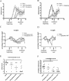
Figure 1-6916
Figure 1-6916
Daily intraperitoneal administration of CNTF reduces EAE symptoms in mice. A: Mice were immunized with MOG(35-55) peptide and complete Freund’s adjuvant on day 0. From days 8 to 24 mice were either treated with 60 μg/kg/day CNTF (n = 7) (black circles), 180 μg/kg/day CNTF (n = 8) (white triangles), or saline (n = 8) (white squares). Treatment with 180 μg/kg/day significantly improved the clinical course during the time of treatment. B: Intraperitoneal injection of 1.2 mg/kg/day CNTF (n = 11) (black diamonds) led to a further improvement of the clinical symptoms compared to saline-treated mice (n = 5) (white squares). C: Intraperitoneal injection of 60 or 180 μg/kg/day CNTF modified the time course of weight loss in mice with EAE, but minimal body weight did not change significantly between CNTF- and saline-injected mice. D: Treatment with 1.2 mg/kg/day induced severe loss of body weight in some mice. These mice were sacrificed at day 23. Cessation of CNTF treatment at day 24 led to increase in body weight. E: Treatment with 1.2 mg/kg/day CNTF led to a significant improvement of the maximal clinical score during the treatment period compared to saline-injected mice (P = 0.02). F: Treatment with 180 μg/kg/day and 1.2 mg/kg/day significantly reduced the cumulative disease score during treatment period compared to saline-injected mice (180 μg/kg/day, P = 0.0112; 1.2 mg/kg/day, P = 0.0016). The graphs in E and F show the median scores. One sacrificed because of severe EAE and two sacrificed because of severe weight loss.
Figure 2-6916
Figure 2-6916
CNTF does not modify T-cell proliferation and cytokine mRNA levels in response to immunization with MOG peptide. A: Lymph nodes were collected from mice immunized with MOG plus complete Freund’s adjuvant and treated with 180 μg/kg/day (n = 8), 1.2 mg/kg/day CNTF (n = 7), or saline (n = 11) at days 15 to 22 after immunization. Proliferative response of isolated LN cells was assayed in vitro. Stimulation with 50 μg of MOG(35-55) peptide led to a marked MOG-dependent proliferation that was not altered by CNTF treatment. B and C: Cytokine mRNA levels were assessed by real-time PCR analysis of RNA isolated from lymph nodes or spleen. The results for 180 μg/kg/day (n = 9) and 1.2 mg/kg/day CNTF (n = 2) were derived from two separate experiments. Results are shown within one graph with the appropriate saline control groups (n = 9 or 6, respectively). B: The levels of IFN-γ mRNA did not differ significantly between CNTF-treated and saline-injected mice. C: Mice treated with 1.2 mg/kg/day CNTF had significantly higher levels of IL-10 mRNA. D–F: Levels of IL-2, IFN-γ, and IL-5 protein in the supernatants of lymph node cell cultures from mice treated with saline (n = 8) or 1.2 mg/kg/day CNTF (n = 6) were measured by ELISA. No significant differences were detected.
Figure 3-6916
Figure 3-6916
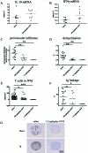
CNTF reduces the immune response in the spinal cord. Levels of mRNA for IL-10 and IFN-γ, number of perivascular infiltrates, extent of demyelination, number of T cells in unaffected white matter, and permeability of the BBB for Ig were analyzed in spinal cord during the acute phase of the disease. A and B: Mice were treated with 180 μg/kg/day CNTF (n = 9) or saline (n = 9) and levels of mRNA for IL-10 and IFN-γ were determined by real-time PCR. In mice treated with 180 μg/kg/day CNTF (n = 9) a trend to lower mRNA levels for IL-10 (P = 0.1513) (A) and significantly lower IFN-γ (P = 0.0135) (B) were observed in spinal cord tissue compared to saline-injected mice (n = 9). C–F: Tissue sections from mice injected with saline (n = 16), 180 μg/kg/day (n = 10) or 1.2 mg/kg/day CNTF (n = 4) were stained with the macrophage marker MAC3, the T-cell marker CD3, the myelin stain LFB-PAS, and anti-Ig for BBB leakage. Mice treated with CNTF showed significantly fewer perivascular infiltrates/cross-section (C), reduced loss of LFB-PAS staining (D), fewer T cells in the apparently unaffected white matter (E), and reduced Ig leakage (F) compared to saline-injected mice. G: Immunohistochemistry for MAC3 and Ig revealed a complete absence of perivascular macrophages/microglia cells and BBB leakage in mice treated with 1.2 mg/kg/day in contrast to saline-injected mice. Arrows indicate perivascular infiltrates [immunohistochemistry for Mac3 (brown) and mouse Ig (brown), counterstained with hematoxylin]. The graphs in A–F show means. In A–D and F, each point represents one animal, in E each point shows the result for a single spinal cord cross-section.
Figure 4-6916
Figure 4-6916
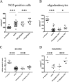
CNTF increases the number of oligodendrocytes and their precursors and decreases axonal and neuronal damage. A–D: To analyze the effects of CNTF treatment in the chronic disease phase, spinal cord tissues from mice in the chronic disease phase [days 28 to 36 after immunization with MOG(35-55) peptide] were studied. The numbers of NG-2-positive cells, oligodendrocytes (PLP-ISH), axons (Bielschowsky’s staining), and neurons (NeuN) in healthy mice or mice treated either with different concentrations of CNTF or saline were determined. The animals from the two experiments shown in Figure 1, A and B, were pooled for this analysis; the saline-treated mice in these two experiments did not show significant differences in the clinical course or different histological parameters. A–C: In saline-treated EAE, the numbers of NG2-positive cells, PLP-positive oligodendrocytes, and axons within the lesions were significantly reduced compared to naive mice (P < 0.001 for all parameters). D: Also the number of NeuN-positive neurons in the ventral horn of the thoracic spinal cord of saline-injected mice with EAE was significantly lower compared to healthy animals without disease (P < 0.001). Treatment with 1.2 mg/kg/day CNTF led to significantly higher numbers of NG-2-positive cells (P = 0.0002) (A), PLP-positive oligodendrocytes (P = 0.012) (B), and axons (P < 0.0001) (C) within the lesions. D: Administration of 1.2 mg/kg/day CNTF prevented completely the reduction in NeuN-positive neurons observed in control EAE mice (P = 0.0006). Notice the different scale of the y axis in D. Each point in A–C represents the result of one spinal cord cross-section, in D each dot represents the result of one animal.
Figure 5-6916
Figure 5-6916
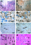
Morphological changes in EAE lesions. Immunohistological analysis of mice in the chronic disease phase (days 28 to 36 after immunization with MOG(35-55) peptide. A: Immunization with MOG(35-55) peptide leads to focal loss of oligodendrocytes and myelin [immunohistochemistry for PLP (red) combined with in situ hybridization for PLP mRNA (blue cytoplasmic signal)]. The arrows mark the border of the lesion. B: EAE lesions are characterized by a clear reduction in the number of axons (same lesion as in A, Bielschowsky’s silver impregnation). C: In EAE lesions cells with morphological characteristics of apoptosis such as condensed and/or fragmented nuclei are found (arrows). D: A proportion of apoptotic cells are oligodendrocytes as identified by immunohistochemistry with CNPase (brown). The apoptotic cell expresses CNPase on the cell surface (arrow). C and D: Immunohistochemistry with anti-CNPase, counterstained with hematoxylin. E and F: Immunohistochemistry for NG2 (brown) reveals a marked up-regulation of NG2-positive cells (arrows) in EAE lesions (F) compared to unaffected white matter (E). G: Within white matter lesions numerous cells with fragmented DNA are observed that stain positive in the TUNEL reaction (left, black arrows). The right panel depicts the adjacent gray matter. No TUNEL-positive neurons are seen in the gray matter (arrows), but one small nonneuronal cell is TUNEL-positive (arrowhead) [TUNEL staining (black) combined with nuclear fast red]. H: In the anterior horns of spinal cord gray matter swollen, weakly eosinophilic neurons were found that lack Nissl bodies (arrows).

Similar articles

Cited by

References

    1. Keegan BM, Noseworthy JH. Multiple sclerosis. Annu Rev Med. 2002;53:285–302. - PubMed
    1. Owens T. The enigma of multiple sclerosis. Curr Opin Neurol. 2003;16:259–265. - PubMed
    1. Prineas JW. The neuropathology of multiple sclerosis. Koetsier JC, editor. Elsevier Science Publishers,; Amsterdam: Demyelinating Diseases. 1985:pp 213–257.
    1. Lassmann H. Springer-Verlag,; Berlin: Comparative Neuropathology of Chronic Experimental Allergic Encephalomyelitis and Multiple Sclerosis. 1983 - PubMed
    1. Bjartmar C, Kidd GJ, Mork S, Rudick R, Trapp BD. Neurological disability correlates with spinal cord axonal loss and reduced N-acetyl aspartate in chronic multiple sclerosis patients. Ann Neurol. 2000;48:893–901. - PubMed

Publication types

MeSH terms

LinkOut - more resources