. 2006 Aug;169(2):682-96.
doi: 10.2353/ajpath.2006.051047.

Androgen receptor remains critical for cell-cycle progression in androgen-independent CWR22 prostate cancer cells

Affiliations

Androgen receptor remains critical for cell-cycle progression in androgen-independent CWR22 prostate cancer cells

Xin Yuan et al. Am J Pathol. 2006 Aug.

Abstract

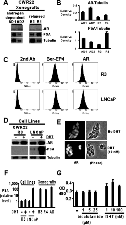
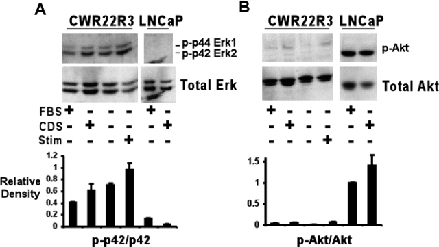
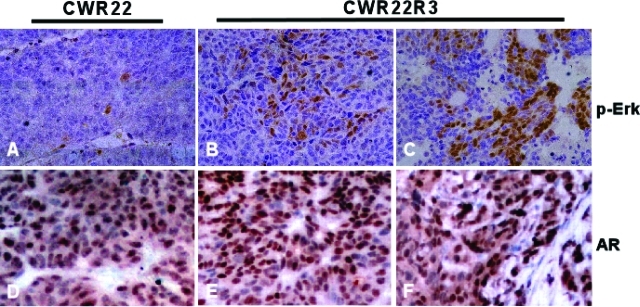
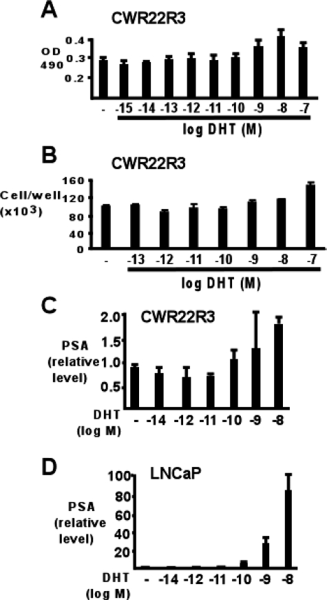
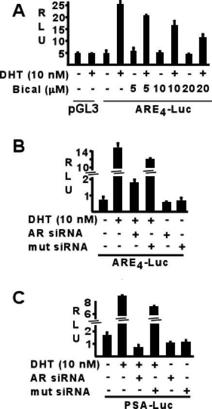
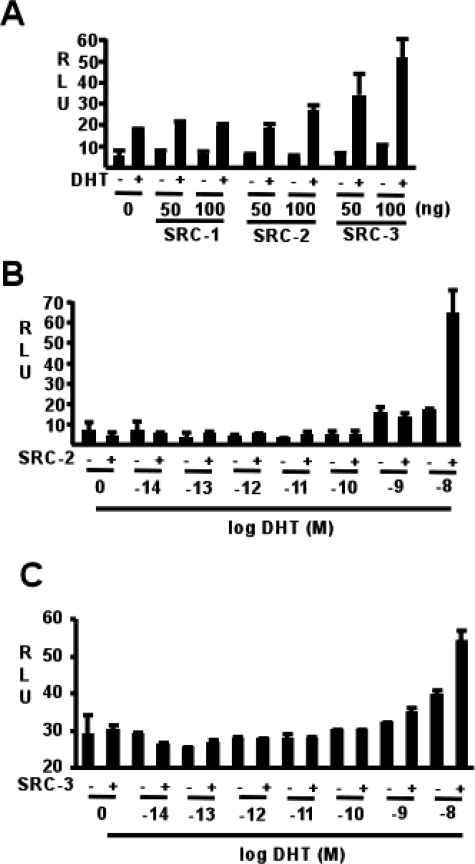
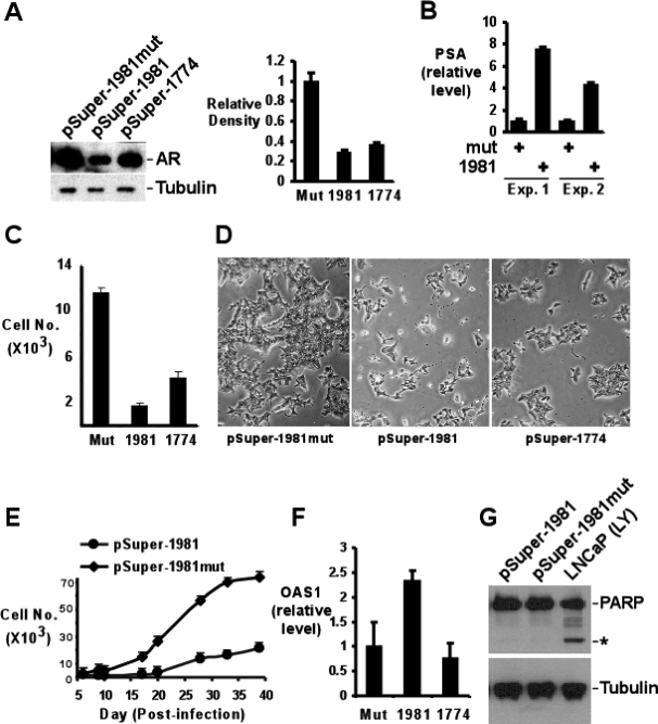
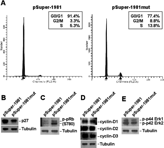
The majority of prostate cancers (PCa) that relapse after androgen deprivation therapy (androgen-independent PCa) continue to express androgen receptor (AR). To study the functional importance of AR in these tumors, we derived androgen-independent CWR22 PCa xenografts in castrated mice and generated a cell line from one of these xenografts (CWR22R3). Similarly to androgen-independent PCa in patients, the relapsed xenografts and cell line expressed AR and were resistant to treatment with bicalutamide. However, expression of the AR-regulated PSA gene in the CWR22R3 cell line was markedly decreased compared to the relapsed xenografts in vivo. Transfections with androgen-regulated reporter genes further indicated that the cells lacked androgen-independent AR transcriptional activity and were not hypersensitive to low androgen concentrations despite constitutive activation of the Erk/MAP kinases. Nonetheless, AR remained essential for androgen-independent growth because retroviral shRNA-mediated AR down-regulation resulted in marked long-term growth suppression. This was associated with increased levels of p27(kip1) and hypophosphorylation of retinoblastoma protein but not with decreases in D-type cyclin levels or MAP kinase activation. These results reveal a potentially critical function of AR in androgen-independent PCa that is distinct from its previously described transcriptional or nontranscriptional functions.


Figures

Figure 1-6930
Figure 1-6930
Characterization of CWR22 xenografts and cell lines. A: Western blot analysis to compare the AR and PSA protein levels in the relapsed and bicalutamide-resistant CWR22 xenografts (relapsed R3, R4) with the levels in two of their androgen-dependent counterparts (androgen-dependent, AD1 and AD2). Twenty μg of protein from each sample were loaded. β-Tubulin was blotted for protein loading control. B: Quantitation of AR and PSA expression. Western blot shown in A was analyzed with the ImageJ program, and the AR and PSA expression in each sample was shown as relative to β-tubulin in each sample. C: Expression of Ber-EP4 and AR in CWR22R3 and LNCaP cells. CWR22R3 cells (R3) were grown in CDS medium. LNCaP cells were grown in RPMI with 10% FBS. No additional DHT was added. Cells were fixed and permeabilized. Cells were subsequently treated with fluorescein isothiocyanate-conjugated anti-mouse antibody alone (second antibody) or with mouse anti-Ber-EP4 (Ber-EP4) or anti-AR (AR) antibodies first, and followed by treating with fluorescein isothiocyanate-conjugated anti-mouse antibody. The labeled cells were analyzed with fluorescence-activated cell sorting. D: Western blot analysis of AR and PSA protein expression in CWR22R3 and LNCaP cell lines. The cells were grown in CDS medium for 3 days and then incubated with (+) or without (−) added 10 nmol/L DHT for an additional 24 hours. The samples were also blotted for α-tubulin for protein loading amount control. E: Immunofluorescence analysis of AR expression in CWR22R3 cells. Cells were grown in CDS medium and incubated without (no DHT) or with 10 nmol/L DHT (DHT) for 24 hours. The left panels showed the staining results by an anti-AR antibody. The right panels showed the phase contrast images of the cells. F: Quantitative real-time RT-PCR analysis of PSA mRNA levels. CWR22R3 (R3) or LNCaP cells were cultured in CDS medium for 48 hours before being changed to CDS medium with (+) or without (−) 10 nmol/L DHT supplement for an additional 24 hours. Total RNA was extracted from the above treated cells as well as from CWR22R3 and R4 xenografts (R3, R4) and an androgen-dependent CWR22 xenograft (AD). The amount of PSA mRNA in each sample is presented as the fold of change (relative level) above the PSA mRNA amount in CWR22R3 cells cultured in CDS medium without DHT supplement. G: Growth of CWR22R3 cells in response to bicalutamide and DHT. Cells were grown in CDS medium alone or with bicalutamide (1 to 25 μmol/L) or DHT (1 to 100 nmol/L) for 48 hours. The vital cells were measured by MTS assays as arbitrary OD units at 490-nm wavelength. Original magnifications, ×400.
Figure 2-6930
Figure 2-6930
Erk activation in the CWR22R3 cell line. CWR22R3 or LNCaP cells were grown in FBS (FBS+) or CDS medium (CDS+) for 24 hours. Additional CWR22R3 cells were grown in serum-free medium (FBS−, CDS−, stim−) for 24 hours, with or without subsequent serum stimulation with CDS medium for 20 minutes (FBS−, CDS−, stim+, or FBS−, CDS−, stim−). The cell lysates were blotted with anti-phospho-Erk (p-p44 Erk1/p-p42 Erk2) and anti-total Erk (total Erk) antibodies (A, top); or anti-phospho-Akt (p-Akt) and anti-total Akt (total Akt) antibodies (B, top). The bottom panels showed the quantification of Erk phosphorylation (A, p-p42/p42) or Akt phosphorylation (B, p-Akt/Akt) by ImageJ analysis of the respective top panels.
Figure 3-6930
Figure 3-6930
Erk activation during the relapse of CWR22R3 xenograft. Immunohistochemical staining with phospho-Erk-specific antibodies (A–C) or anti-AR antibodies (D–E) on CWR22R3 xenograft biopsy samples prepared before castration (A, D), after castration but before the initiation of bicalutamide treatment (B, E), or at the end of bicalutamide treatment (C, F). Original magnifications, ×200.
Figure 4-6930
Figure 4-6930
CWR22R3 growth and PSA expression in response to DHT. A: CWR22R3 growth in response to added DHT. Cells were grown in CDS medium alone or with added DHT (10−15 to 10−7 mol/L) for 48 hours. The vital cells were measured by MTS assays as arbitrary OD units at 490-nm wavelength. B: CWR22R3 were grown in CDS medium alone or with added DHT (10−13 to 10−7 mol/L) for 48 hours and number of cells were directly counted. C and D: Real-time RT-PCR analysis of PSA mRNA expression in CWR22R3 cells (C) and LNCaP (D) grown in CDS medium for 48 hours followed by stimulation with 0, 10−14 to 10−8 mol/L DHT for an additional 24 hours. The PSA levels are presented as the fold change (relative level) of the PSA level in cells without DHT supplement.
Figure 5-6930
Figure 5-6930
AR transcriptional activity on AR-responsive reporters. CWR22R3 cells grown in CDS medium were transfected with ARE-less pGL3/promoter (pGL3) or with AR-responsive element-driven firefly luciferase reporters, ARE4-Luc or PSA-Luc, as well as a pRL-CMV Renilla luciferase reporter. Twenty-four hours after transfection, the culture media were replaced with CDS medium with or without 10 nmol/L DHT for an additional 24 hours. The cells were lysed at the end of the treatment and AR transcriptional activity is presented as relative light units (firefly luciferase over Renilla luciferase activity, RLU). A: The posttransfected cells were co-treated with various concentrations of bicalutamide (Bical) during the last 24 hours. B and C: ARE4-Luc (B) and PSA-Luc (C) were co-transfected with AR-specific (AR siRNA) or control (mut siRNA) siRNAs and cultured with or without DHT during the last 24 hours.
Figure 6-6930
Figure 6-6930
Effect of supplemented SRC proteins on AR transcriptional activity. A: CWR22R3 cells grown in CDS medium were transfected with AR-responsive reporters, ARE4-Luc as well as pRL-CMV Renilla luciferase reporter. Twenty-four hours after transfection, the culture media were replaced with CDS medium with or without 10−8 mol/L DHT for an additional 24 hours. Cells were co-transfected with 0, 50, or 100 ng of SRCs 1 to 3 as indicated. B and C: Cells were co-transfected with 100 ng of SRC-2 (B) or 100 ng of SRC-3 (C) and treated with 0 or 10−14 to 10−8 mol/L DHT during the last 24 hours of the assay. AR transcriptional activity is presented as relative light unit of firefly over Renilla luciferase activity (RLU).
Figure 7-6930
Figure 7-6930
Growth retardation of CWR22R3 cells because of viral-mediated AR down-regulation. A: AR down-regulation by shRNA. CWR22R3 cells were infected with AR-specific (pSuper-1981 or pSuper-1774) or control (pSuper-1981mut) shRNA-generating retroviruses for 2 days. The infected cells were then selected in CDS medium with 2 μg/ml of puromycin for 3 days. The cells were allowed to recover in puromycin-free medium for an additional 2 days before lysis. The cell extracts were blotted for AR and α-tubulin (tubulin) (left). The right panel quantified the AR levels, relative to the respective α-tubulin level in each sample by analyzing the leftpanel blot using the ImageJ program. B: PSA gene expression after AR down-regulation. In two independent experiments (Exp.1 and 2), CWR22R3 cells were infected with pSuper-1981mut (mut) or pSuper-1981(1981), and the infected cells were selected with puromycin as in A. Total RNA was isolated at days 7 (Exp. 1) or 8 (Exp. 2) after infection. Quantitative real-time RT-PCR analysis of PSA mRNA levels was presented as the relative fold of change (relative level) compared to the PSA mRNA level in mut infected cells. C: Cells counted at 9 days after infection with AR-specific (1981 or 1774) or control (mut) shRNA-generating retroviruses and after puromycin selection. D: Phase contrast images of CWR22R3 cells at 9 days after infection with AR-specific (pSuper-1981 or pSuper-1774) or control (pSuper-1981mut) shRNA-generating retroviruses and after puromycin selection. E: Viral-infected and puromycin-selected CWR22R3 cells (2 × 10³) at 5 days after infection were plated in each 24-well well and grown in CDS medium. The cell growth was followed by counting cells at indicated days after infection. F: OAS1 induction in CWR22R3 cells at 11 days after infection infected with AR-specific (1981 or 1774) or control (mut) shRNA-generating retroviruses and after puromycin selection. The induction was measured by quantitative real-time RT-PCR and presented as relative fold of change (relative level) compared to that in mut-infected cells. G: Cell extracts from CWR22R3 cells at 3 days after infection without puromycin selection were blotted for PARP and α-tubulin (tubulin). An asterisk indicates the cleaved PARP products. Original magnifications, ×200.
Figure 8-6930
Figure 8-6930
Cell-cycle arrest by AR down-regulation. Puromycin-selected CWR22R3 cells were collected at day 7 after infection with AR-specific (pSuper-1981) or control (pSuper-1981mut) shRNA retroviruses and after puromycin selection. A: Cell-cycle analysis by propidium iodide labeling and flow cytometry. The percentages of cells in each cell-cycle phase are listed. Cell lysates were blotted for p27kip1 (p27) (B), phospho-retinoblastoma protein (p-pRB, S780) (C), cyclin D1–3 (D), and phospho-p44 Erk1 and phospho-p42 Erk2 (p-p44 Erk1 and p-p42 Erk2) (E). α-Tubulin (tubulin) was also blotted to indicate comparable protein loading in each sample.

Similar articles

Cited by

References

    1. Gelmann EP. Molecular biology of the androgen receptor. J Clin Oncol. 2002;20:3001–3015. - PubMed
    1. Feldman BJ, Feldman D. The development of androgen-independent prostate cancer. Nature Rev Cancer. 2001;1:34–45. - PubMed
    1. Balk SP. Androgen receptor as a target in androgen-independent prostate cancer. Urology. 2002;60:132–138. - PubMed
    1. Joyce R, Fenton MA, Rode P, Constantine M, Gaynes L, Kolvenbag G, DeWolf W, Balk S, Taplin ME, Bubley GJ. High dose bicalutamide for androgen independent prostate cancer: effect of prior hormonal therapy. J Urol. 1998;159:149–153. - PubMed
    1. Small EJ, Halabi S, Dawson NA, Stadler WM, Rini BI, Picus J, Gable P, Torti FM, Kaplan E, Vogelzang NJ. Antiandrogen withdrawal alone or in combination with ketoconazole in androgen-independent prostate cancer patients: a phase III trial (CALGB 9583). J Clin Oncol. 2004;22:1025–1033. - PubMed

Publication types

MeSH terms