

STAT-6-mediated control of P-selectin by substance P and interleukin-4 in human dermal endothelial cells
- PMID: 16877367
- PMCID: PMC1698799
- DOI: 10.2353/ajpath.2006.051211
STAT-6-mediated control of P-selectin by substance P and interleukin-4 in human dermal endothelial cells
Abstract
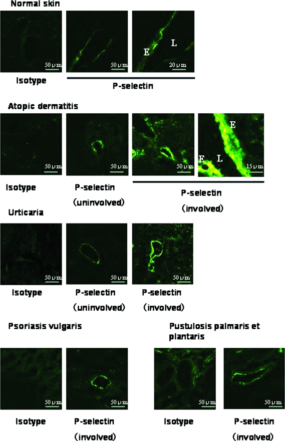
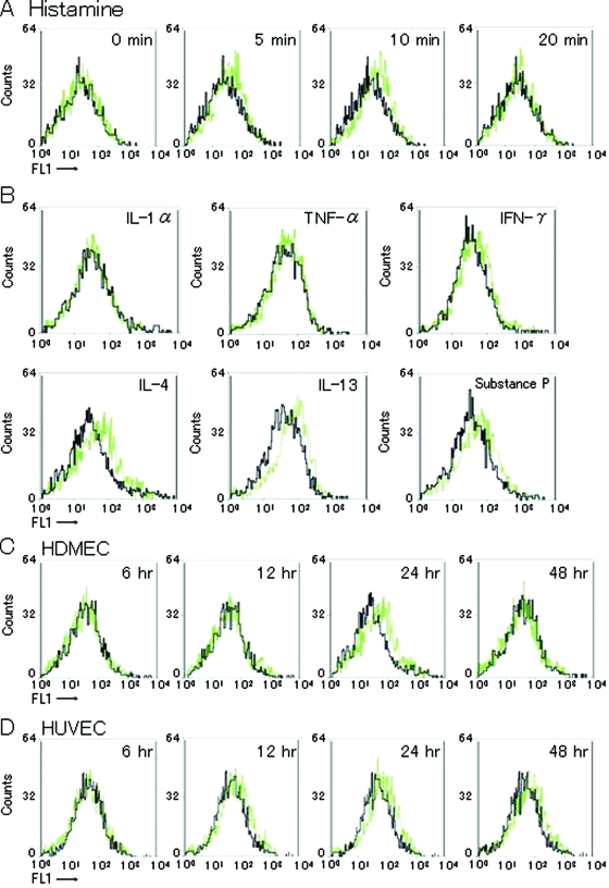
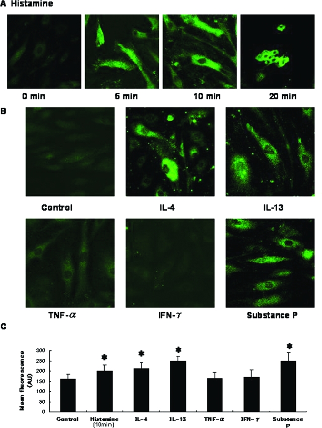
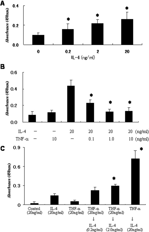
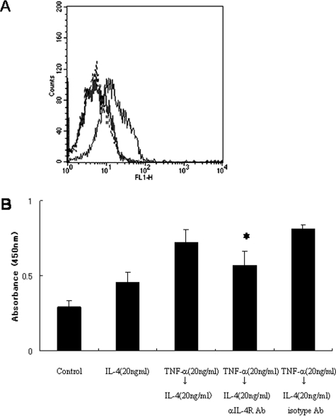
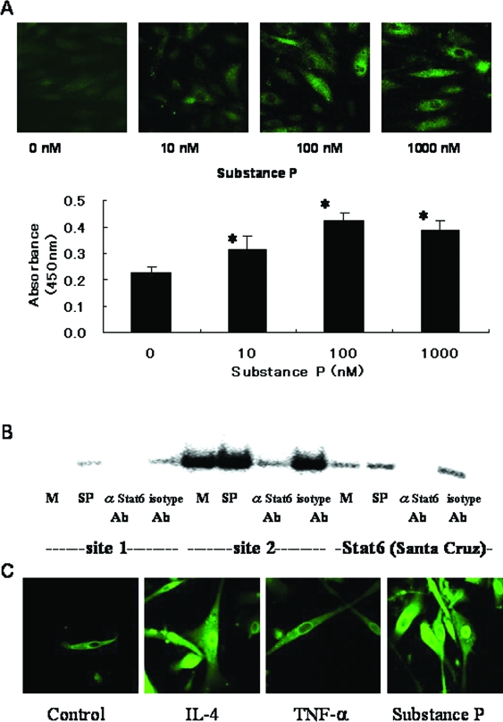
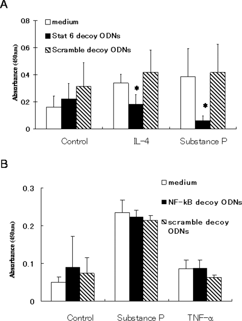
P-Selectin expressed on endothelial cells contributes to acute and chronic inflammation by promoting leukocyte tethering/rolling. Despite increasing evidence of P-selectin expression on human umbilical vein endothelial cells in vitro, the regulatory mechanisms of P-selectin expression on dermal endothelial cells in skin diseases are not fully understood. Here, we demonstrate increased expression of P-selectin in dermal vessels of regional skin in urticaria and atopic dermatitis. The present in vitro analyses with human dermal microvascular endothelial cells (HDMECs) revealed that histamine rapidly induced P-selectin expression. Interleukin (IL)-4 and IL-13 induced prolonged expression of surface P-selectin by HDMECs. A combination of tumor necrosis factor-alpha and IL-4 inhibited P-selectin expression. Pretreatment of HDMECs with tumor necrosis factor-alpha followed by incubation with IL-4 markedly increased P-selectin expression. Notably, incubation with substance P alone induced prolonged P-selectin expression. Activation of STAT6 appears to be a key factor in P-selectin expression induced by substance P and IL-4 because treatment with STAT6 decoy oligodeoxynucleotides significantly inhibited P-selectin expression. The present results indicate that novel, complex mechanisms are involved in endothelial P-selectin expression in the skin. STAT6 in dermal endothelial cells appears to be a potent target for controlling cellular infiltrate in allergic and/or neuroinflammatory skin diseases.
Figures
Similar articles
-
Immature mast cells exhibit rolling and adhesion to endothelial cells and subsequent diapedesis triggered by E- and P-selectin, VCAM-1 and PECAM-1.Exp Dermatol. 2010 May;19(5):424-34. doi: 10.1111/j.1600-0625.2010.01073.x. Exp Dermatol. 2010. PMID: 20507363
-
IL-4 and IL-13 downregulate rolling adhesion of leukocytes to IL-1 or TNF-alpha-activated endothelial cells by limiting the interval of E-selectin expression.Cytokine. 1998 Jun;10(6):395-403. doi: 10.1006/cyto.1997.0308. Cytokine. 1998. PMID: 9632524
-
UV radiation induces the release of angiopoietin-2 from dermal microvascular endothelial cells.Exp Dermatol. 2012 Feb;21(2):147-53. doi: 10.1111/j.1600-0625.2011.01416.x. Epub 2011 Dec 6. Exp Dermatol. 2012. PMID: 22142364 Clinical Trial.
-
Stat6 activation is essential for interleukin-4 induction of P-selectin transcription in human umbilical vein endothelial cells.Arterioscler Thromb Vasc Biol. 1999 Jun;19(6):1421-9. doi: 10.1161/01.atv.19.6.1421. Arterioscler Thromb Vasc Biol. 1999. PMID: 10364072
-
Regulation of substance P mRNA expression in human dermal microvascular endothelial cells.Clin Exp Rheumatol. 2004 Jan-Feb;22(3 Suppl 33):S24-7. Clin Exp Rheumatol. 2004. PMID: 15344593 Review.
Cited by
-
Novel functional aspect of antihistamines: the impact of bepotastine besilate on substance p-induced events.J Allergy (Cairo). 2009;2009:853687. doi: 10.1155/2009/853687. Epub 2009 Jun 21. J Allergy (Cairo). 2009. PMID: 20975801 Free PMC article.
-
Mucosal neuroimmune mechanisms in gastro-oesophageal reflux disease (GORD) pathogenesis.J Gastroenterol. 2024 Mar;59(3):165-178. doi: 10.1007/s00535-023-02065-9. Epub 2024 Jan 14. J Gastroenterol. 2024. PMID: 38221552 Free PMC article. Review.
-
Differential gene expression of primary cultured lymphatic and blood vascular endothelial cells.Neoplasia. 2007 Dec;9(12):1038-45. doi: 10.1593/neo.07643. Neoplasia. 2007. PMID: 18084611 Free PMC article.
-
Genome-wide approaches reveal functional interleukin-4-inducible STAT6 binding to the vascular cell adhesion molecule 1 promoter.Mol Cell Biol. 2011 Jun;31(11):2196-209. doi: 10.1128/MCB.01430-10. Epub 2011 Apr 4. Mol Cell Biol. 2011. PMID: 21464207 Free PMC article.
-
P-Selectin Expressed by a Human SELP Transgene Is Atherogenic in Apolipoprotein E-Deficient Mice.Arterioscler Thromb Vasc Biol. 2016 Jun;36(6):1114-21. doi: 10.1161/ATVBAHA.116.307437. Epub 2016 Apr 21. Arterioscler Thromb Vasc Biol. 2016. PMID: 27102967 Free PMC article.
References
-
- Catalina MD, Estess P, Siegelman MH. Selective requirements for leukocyte adhesion molecules in models of acute and chronic cutaneous inflammation: participation of E- and P- but not L-selectin. Blood. 1999;93:580–589. - PubMed
-
- Kitayama J, Fuhlbrigge RC, Puri KD, Springer TA. P-selectin, L-selectin, and α4 integrin have distinct roles in eosinophil tethering and arrest on vascular endothelial cells under physiological flow conditions. J Immunol. 1997;159:3929–3939. - PubMed
-
- Patel KD, McEver RP. Comparison of tethering and rolling of eosinophils and neutrophils through selectins and P-selectin glycoprotein ligand-1. J Immunol. 1997;159:4555–4565. - PubMed
-
- Satoh T, Kaneko M, Wu M-H, Yokozeki H, Nishioka K. Contribution of selectin ligands to eosinophil recruitment into the skin of patients with atopic dermatitis. Eur J Immunol. 2002;32:1274–1281. - PubMed
Publication types
MeSH terms
Substances
LinkOut - more resources
Full Text Sources
Research Materials
Miscellaneous
