. 2006 Aug;47(8):3450-60.
doi: 10.1167/iovs.05-1208.

Lens-specific expression of TGF-beta induces anterior subcapsular cataract formation in the absence of Smad3

Affiliations

Lens-specific expression of TGF-beta induces anterior subcapsular cataract formation in the absence of Smad3

Alice Banh et al. Invest Ophthalmol Vis Sci. 2006 Aug.

Abstract

Purpose: Smad3, a mediator of TGF-beta signaling has been shown to be involved in the epithelial-to-mesenchymal transformation (EMT) of lens epithelial cells in a lens injury model. In this study, the role of Smad3 in anterior subcapsular cataract (ASC) formation was investigated in a transgenic TGF-beta/Smad3 knockout mouse model.

Methods: TGF-beta1 transgenic mice (containing a human TGF-beta1 cDNA construct expressed under the alphaA-crystallin promoter) were bred with mice on a Smad3-null background to generate mice with the following genotypes: TGF-beta1/Smad3(-/-) (null), TGF-beta1/Smad3(+/-), TGF-beta1/Smad3(+/+), and nontransgenic/Smad3(+/+). Lenses from mice of each genotype were dissected and prepared for histologic or optical analyses.

Results: All transgenic TGF-beta1 lenses demonstrated subcapsular plaque formation and EMT as indicated by the expression of alpha-smooth muscle actin. However, the sizes of the plaques were reduced in the TGF-beta1/Smad3(-/-) lenses, as was the level of type IV collagen deposition when compared with TGF-beta1/Smad3(+/-) and TGF-beta1/Smad3(+/+) lenses. An increased number of apoptotic figures was also observed in the plaques of the TGF-beta1/Smad3(-/-) lenses compared with TGF-beta1/Smad3(+/+) littermates.

Conclusions: Lens-specific expression of TGF-beta1 induced ASC formation in the absence of the Smad3 signaling mediator, suggests that alternative TGF-beta-signaling pathways participate in this ocular fibrotic model.


Figures
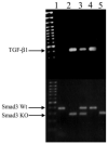
Figure 1
Figure 1
Genotyping for TGF-β1/Smad3 knockout mice. Top gel: PCR results for the TGF-β1 (300 bp) transgene; bottom gel: PCR results for the Smad3 wild-type (Wt, 400 bp) and Smad3 knockout (KO, 250 bp) alleles. The mice had the following genotypes: wild type (lane 1), TGF-β1/Smad3-/- (lane 2), TGF-β1/Smad3+/- (lane 3), TGF-β1/Smad3+/+ (lane 4) and Smad3-/- (lane 5).
FIGURE 2
FIGURE 2
Phosphorylated Smad3. Western blot analysis using an anti-pSmad3/Smad1 antibody for the detection of pSmad3 (58 kDa) protein expression in lens extracts. The antibody cross-reacts with phosphorylated Smad1 (65 kDa) protein, which served as an internal positive control. The lanes show the expression of phospho-Smad3 protein in TGF-β1/Smad3+/+ (lane 1), wild-type (lane 2), TGFβ1/Smad3-/- (lane 3), Smad3-/- (lane 4), and TGF-β1/Smad3+/- (lane 5) mouse lenses. Both TGF-β1/Smad3+/+ and TGF-β1/Smad3+/- lenses showed the presence of pSmad3, whereas wild-type and Smad3-null (TGF-β1/Smad3-/- and Smad3-/-) lenses did not.
FIGURE 3
FIGURE 3
Histologic analysis of the TGF-β1/Smad3 lenses. Wild-type (A, E, I), TGF-β1/Smad3+/+ (B, F, J), TGF-β1/Smad3-/- (C, G, K), and a Smad3-/- (D, H, L) lenses are shown. Boxes: areas in higher-magnification images. The expression of the TGF-β1 transgene induced subcapsular plaque formation in the TGF-β1/Smad3+/+ and TGF-β1/Smad3-/- lenses (F, G, small arrows). The subcapsular plaques in TGF-β1/Smad3-/- lenses were smaller than those in the TGF-β1/Smad3+/+ lenses. Both the wild-type and Smad3-/- lenses showed normal morphology of the lens epithelium. The scale bars: (A-D) 400 μm; (E-H) 100 μm; (I-L) 50 μm.
FIGURE 4
FIGURE 4
Vacuole formation in the TGF-β1/Smad3 lenses. The posterior lens cortex of wild-type (A), TGF-β1/Smad3+/+ (B), and TGF-β1/Smad3-/- (C) lenses are shown. The expression of TGF-β1 induced nucleation and vacuole formation in the TGF-β1/Smad3+/+ lenses. Both the wild-type and TGF-β/Smad3-/- lenses showed normal morphology of the posterior lens cortex. Scale bar, 200 μm.
FIGURE 5
FIGURE 5
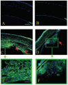
Immunohistochemical analysis of α-SMA expression (FITC). Wild-type (A), Smad3-/- (B), TGF-β1/Smad3+/+ (C, E), and TGF-β1/Smad3-/- (D, F) lenses are shown. Red arrows: subcapsular plaques. Expression of α- SMA was detected in the subcapsular plaques of the TGF-β1/Smad3+/+ and TGF-β1/Smad3-/- lenses. However, there was less α-SMA immunoreactivity detected in TGF-β1/Smad3-/- lenses. Both the wild-type and Smad3-/- lenses showed no expression of α-SMA in the lens and normal expression in the iris (positive control). Scale bars: (A-D) 100 μm; (E, F) 50 μm.
FIGURE 6
FIGURE 6
α-Smooth muscle actin protein expression. Western blot analysis using an anti-α-SMA antibody for the detection of α-SMA (42 kDa) protein expression in lens extracts. The membrane was stripped and reprobed for β-actin, which served as a loading control. Lanes 1 to 5: α-SMA and β-actin protein expression in wild-type, Smad3-/-, TGF-β1/Smad3+/-, TGF-β1/Smad3+/+, and TGFβ1/Smad3-/- lenses, respectively. The β-actin signal shows that equal amounts of protein were loaded in all lanes. TGF-β1/Smad3+/+, TGF-β1/Smad3+/-, and TGFβ1/Smad3-/- lenses showed expression of α-SMA, whereas wild-type and Smad3-/- lenses did not. The TGFβ1/Smad3-/- lenses showed reduced levels of α-SMA compared with both the TGF-β1/Smad3+/+ and TGF-β1/Smad3+/- lenses.
FIGURE 7
FIGURE 7
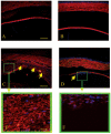
Immunohistochemical analysis of fibronectin expression (TRITC). Wild-type (A), Smad3-/- (B), TGF-β1/Smad3+/+ (C, E) and TGF-β1/Smad3-/- (D, F) lenses are shown. Yellow arrows: location of subcapsular plaques. There was detectable expression of fibronectin in the subcapsular plaques of the TGF-β1/Smad3+/+ and TGF-β1/Smad3-/- lenses. The intensity of fibronectin immunoreactivity in the subcapsular plaques of TGF-β1/Smad3-/- lenses was reduced when compared to the TGF-β1/Smad3+/+ lenses. Both the wild-type and Smad3-/- lenses showed normal expression of fibronectin in the lens capsule. Scale bars: (A-D) 100 μm; (E, F) 50 μm.
FIGURE 8
FIGURE 8
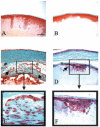
Mason’s trichrome staining for collagen. Wild-type (A), Smad3-/- (B), TGF-β1/Smad3+/+ (C, E), and TGF-β1/Smad3-/- (D, F) lenses are shown. Arrows: subcapsular plaques; (*) collagen expression in the lens capsules. The expression of TGF-β1 increased collagen deposition in the TGF-β1/Smad3+/+ and TGF-β1/Smad3-/- plaques. However, the collagen deposition in the subcapsular plaques of TGF-β1/Smad3-/- lenses was substantially reduced compared with the TGF-β1/Smad3+/+ lenses. Both the wild-type and Smad3-/- lenses showed normal collagen expression in the lens capsule. Scale bars: (A-D) 100 μm; (E, F) 50 μm.
FIGURE 9
FIGURE 9
Immunohistochemical analysis of collagen type I expression (FITC). Wild-type (A), Smad3-/- (B), TGF-β1/Smad3+/+ (C, E), and TGF-β1/Smad3-/- (D, F) lenses are shown. Arrows: subcapsular plaques. Expression of collagen type I was detected in the subcapsular plaques of the TGF-β1/Smad3+/+ and TGF-β1/Smad3-/- lenses. There was no observable difference in collagen I expression between the TGF-β1/Smad3-/- and TGF-β1/Smad3+/+ lenses. Both the wild-type and Smad3-/- lenses show no expression of collagen type I in the lens. Scale bars: (A-D) 100 μm; (E, F) 50 μm.
FIGURE 10
FIGURE 10
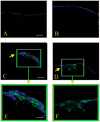
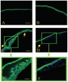
Immunohistochemical analysis of collagen type IV expression (FITC). Wild-type (A), Smad3-/- (B), TGF-β1/Smad3+/+ (C, E), and TGF-β1/Smad3-/- (D, F) lenses are shown. Arrows: subcapsular plaques. Expression of collagen type IV was detected in the subcapsular plaques of the TGF-β1/Smad3+/+ and TGF-β1/Smad3-/- lenses. There was considerable reduction in collagen IV immunoreactivity in the TGF-β1/Smad3-/- lenses when compared with the TGF-β1/Smad3+/+ lenses. Both the wild-type and Smad3-/- lenses showed collagen type IV in the lens capsule. Scale bars: (A-D) 100 μm; (E, F) 50 μm.
FIGURE 11
FIGURE 11
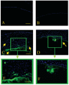
Immunohistochemical analysis of β-crystallin expression (FITC). Shown are wild-type (A), Smad3-/- (B), TGF-β1/Smad3+/+ (C, E), and TGF-β1/Smad3-/- (D, F) mouse lenses. Arrows: subcapsular plaques. Expression of β-crystallin was detected in the posterior aspect (abutting the lens fiber cell mass) of the subcapsular plaques in both the TGF-β1/Smad3+/+ and TGF-β1/Smad3-/- lenses. Both the wild-type and Smad3-/- lenses showed no expression of β-crystallin in the lens epithelium. Scale bars: (A-D) 100 μm; (E, F) 50 μm.
FIGURE 12
FIGURE 12
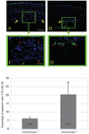
TUNEL staining of apoptotic nuclei (FITC) in TGF-β1/Smad3 lenses. Shown are TGF-β1/Smad3+/+ (A, C) and TGF-β1/Smad3-/- (B, D) mouse lenses. Arrows: subcapsular plaques. The TGF-β1/Smad3+/+ lenses showed a few TUNEL-positive nuclei located in the anterior and posterior regions of the plaques (A, C). The TGF-β1/Smad3-/- lenses exhibited more TUNEL-positive nuclei. The bar graph represents the percentage of apoptotic nuclei per 150 cells. *The TGF-β1/Smad3-/- lenses had significantly more apoptotic nuclei (20.3% ± 5.8%) than did the TGF-β1/Smad3+/+ (6.1% ± 1.1%) lenses (unpaired Student’s t-test: P ≤ 0.05). Scale bars: 100 μm(A, B); 50 μm (C, D).
FIGURE 13
FIGURE 13
Optical effects of subcapsular cataract formation in mouse lenses. Data are the BVD error variability (mm ± SEM) in wild-type, TGF-β1/Smad3+/+, TGF-β1/Smad3+/-, and TGF-β1/Smad3-/- mouse lenses. An increased in BVD error signifies a decrease in sharpness of light focus through the lens. +Statistical analysis (ANOVA: P ≤ 0.05) shows that the TGF-β1/Smad3+/+ and TGF-β1/Smad3+/- lenses had the greatest BVD errors (0.531 ± 0.071 and 0.486 ± 0.040 mm, respectively) when compared to the wild-type (0.067 ± 0.002 mm) lenses. *TGF-β1/Smad3-/- (0.099 ± 0.005 mm) lenses also show a significantly greater BVD error when compared to the wild-type lenses. However, the BVD error of TGF-β1/Smad3-/- lenses is significantly lower than both TGF-β1/Smad3+/+ and TGF-β1/Smad3+/- lenses.
FIGURE 14
FIGURE 14
TGF-β induced Smad-dependent and -independent signaling pathways. On ligand binding, type I and type II receptors are activated, and phosphorylation of R-Smads (Smad2/3) occurs. Phosphorylated R-Smads form heterotrimeric complexes with co-Smad (Smad 4) and translocate to the nucleus. The Smad complexes interact with other transcription factors (TF) at DNA sequence-specific binding sites (transcription factor binding element [TBE] Smad binding element [SBE]) to regulate gene expression. Smad7 is an inhibitory Smad that prevents receptor activation of R-Smads. Smad3 phosphorylation is prevented in Smad3-null mice, and so TGF-β-induced responses are presumed to occur through activation of other TGF-β-induced signaling pathways. TGF-β also induces activation of the MAPK pathways (JNK, p38 and ERK1, 2) through the upstream mediators RhoA, Ras, and TAK1. Additional pathways involving PI3K have been shown to mediate EMT. Modified with permission from Roberts AB, Derynck R. Meeting Report: Signaling Schemes for TGF-β. Sci. STKE 2001;pe43. http://stke.sciencemag.org/cgi/content/full/OC sigtrans;2001/113/pe43. © 2001 AAAS.

Similar articles

Cited by

References

    1. Srinivasan Y, Lovicu FJ, Overbeek PA. Lens-specific expression of transforming growth factor beta1 in transgenic mice causes anterior subcapsular cataracts. J Clin Invest. 1998;101:625–634. - PMC - PubMed
    1. Yang X, Letterio JJ, Lechleider RJ, et al. Targeted disruption of SMAD3 results in impaired mucosal immunity and diminished T cell responsiveness to TGF-beta. EMBO J. 1999;18:1280–1291. - PMC - PubMed
    1. Kurisaki A, Kose S, Yoneda Y, Heldin CH, Moustakas A. Transforming growth factor-beta induces nuclear import of Smad3 in an importin-beta1 and Ran-dependent manner. Mol Biol Cell. 2001;12:1079–1091. - PMC - PubMed
    1. Qing J, Zhang Y, Derynck R. Structural and functional characterization of the transforming growth factor-beta-induced Smad3/c-Jun transcriptional cooperativity. J Biol Chem. 2000;275:38802–38812. - PubMed
    1. Dunker N, Krieglstein K. Reduced programmed cell death in the retina and defects in lens and cornea of Tgfbeta2(-/-) Tgf-beta3(-/-) double-deficient mice. Cell Tissue Res. 2003;313:1–10. - PubMed

Publication types

MeSH terms