. 2006 Aug;18(8):1961-74.
doi: 10.1105/tpc.106.041269. Epub 2006 Jul 28.

The accumulation of oleosins determines the size of seed oilbodies in Arabidopsis

Affiliations

The accumulation of oleosins determines the size of seed oilbodies in Arabidopsis

Rodrigo M P Siloto et al. Plant Cell. 2006 Aug.

Abstract

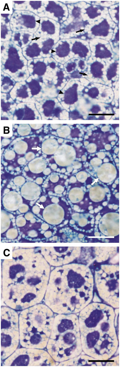
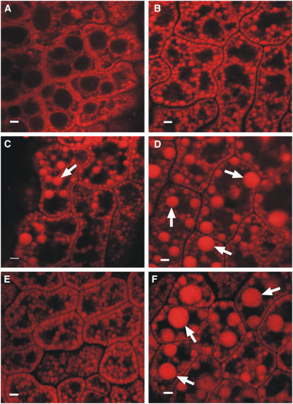
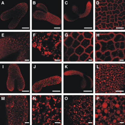
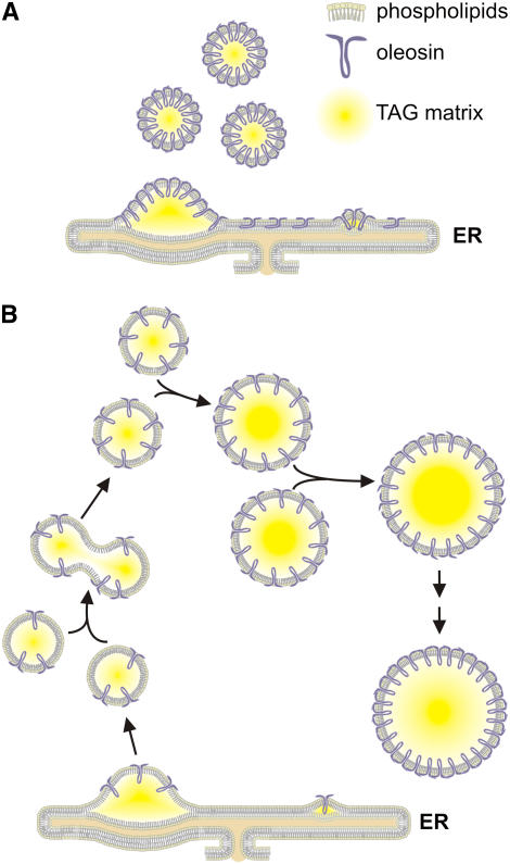
We investigated the role of the oilbody proteins in developing and germinating Arabidopsis thaliana seeds. Seed oilbodies are simple organelles comprising a matrix of triacylglycerol surrounded by a phospholipid monolayer embedded and covered with unique proteins called oleosins. Indirect observations have suggested that oleosins maintain oilbodies as small single units preventing their coalescence during seed desiccation. To understand the role of oleosins during seed development or germination, we created lines of Arabidopsis in which a major oleosin is ablated or severely attenuated. This was achieved using RNA interference techniques and through the use of a T-DNA insertional event, which appears to interrupt the major (18 kD) seed oleosin gene of Arabidopsis and results in ablation of expression. Oleosin suppression resulted in an aberrant phenotype of embryo cells that contain unusually large oilbodies that are not normally observed in seeds. Changes in the size of oilbodies caused disruption of storage organelles, altering accumulation of lipids and proteins and causing delay in germination. The aberrant phenotypes were reversed by reintroducing a recombinant oleosin. Based on this direct evidence, we have shown that oleosins are important proteins in seed tissue for controlling oilbody structure and lipid accumulation.


Figures

Figure 1.
Figure 1.
Suppression of Oleosins in Arabidopsis. (A) Scale diagram of the constructs used to suppress OLEO1 using RNAi methods. The Antisense, Hairpin, and Loop cassettes are shown. (B) Scale diagram of the insertion of a T-DNA element into OLEO1 and OLEO2 genes (lines KnockOLEO1 and KnockOLEO2, respectively). Each gene has two exons (thick line) and one intron (thin line). Both T-DNA insertions are located in the middle of the second exon. (C) SDS-PAGE profile of oilbody-associated proteins in different plants. The first lane (L) contains the protein ladder (Benchmark; Invitrogen). The second lane contains the oilbody-associated proteins from wild-type (C24) plants. Lanes numbered 1 to 3 contain the oilbody-associated proteins from SupOLEO1-Loop, SupOLEO1-Hairpin, and SupOLEO1-Antisense plants, respectively. Lanes 4 and 5 contain the oilbody-associated proteins from KnockOLEO1 and KnockOLEO2 lines, respectively. The positions of the three most abundant oleosin isoforms are indicated by arrows.
Figure 2.
Figure 2.
Phenotype of Arabidopsis Mature Embryos. Bright-field microscopy of osmicated embryos thin-sectioned and stained with toluidine blue. Bars = 10 μm. (A) The wild type. Black arrows indicate oilbodies; arrowheads indicate protein bodies. (B) SupOLEO1-Loop. White arrows indicate larger oilbodies. (C) Plant segregated from SupOLEO1-Loop (null).
Figure 3.
Figure 3.
In Vivo Analysis of Oilbody Morphology. Confocal sections of living mature Arabidopsis embryos stained with Nile red. Large oilbodies in (C), (D), and (F) are shown by arrows. Bars = 2 μm. (A) Wild-type plant. (B) SupOLEO1-Antisense plant. (C) SupOLEO1-Hairpin plant. (D) SupOLEO1-Loop plant. (E) KnockOLEO2 plant. (F) KnockOLEO1 plant.
Figure 4.
Figure 4.
Introduction of a Recombinant Oleosin to Rescue the Oleosin-Deficient Phenotype. SDS-PAGE profiles of oilbody-associated proteins are shown in left panels and confocal sections of mature embryos in right panels. (A) SupOLEO1-Loop plant. OLEO1 polypeptide is indicated by the black arrow. White arrows show large oilbodies. Bar = 5 μm. (B) MaizeOle line. OLEO1 and OLE16 are indicated by the black arrows. Bar = 8 μm. (C) Progeny from crossing between SupOLEO1-Loop and MaizeOle plants. OLEO1 and OLE16 are indicated by the black arrows. Bar = 5 μm.
Figure 5.
Figure 5.
Comparison of Germination Frequency between Wild-Type and SupOLEO1-Loop Plants in Various Conditions. Arabidopsis seeds were germinated in different conditions of light and sucrose availability. (A) Wet filter paper; light. (B) Wet filter paper; stratified seeds; light. (C) Half-strength Murashige and Skoog (MS) medium + sucrose; light. (D) Half-strength MS medium + sucrose; light. (E) Half-strength MS medium − sucrose; dark. (F) Half-strength MS medium + sucrose; dark.
Figure 6.
Figure 6.
Fatty Acid Profiles in Wild-Type, Nulls, and Oleosin-Suppressed Seeds of Arabidopsis. (A) Comparison of major fatty acids (FA) in TAG in wild-type Columbia and oleosin T-DNA insertional knockouts of OLEO1 and OLEO2 in a Columbia background. (B) Comparison of C24 and segregating nulls from the oleosin-suppressed line SupOLEO1-loop.
Figure 7.
Figure 7.
Fate of Oilbodies during Embryo Development. Arabidopsis embryos were collected at different stages of development and stained with Nile red. Bars = 40 μm in (A) and (I), 60 μm in (B) and (J), 150 μm in (C) and (K), 20 μm in (D) and (L), and 5 μm in (E) to (H) and (M) to (P). (A) to (D) Confocal sections of wild-type Arabidopsis embryos at late-heart stage, torpedo stage, walking-stick stage, and mature seed, respectively. (E) to (H) Same as (A) to (D) in higher magnification. (I) to (L) Confocal sections of KnockOLEO1 Arabidopsis embryos at late-heart stage, torpedo stage, walking-stick stage, and mature seed, respectively. (M) to (P) Same as (I) to (L) in higher magnification.
Figure 8.
Figure 8.
Fate of Oilbodies during Seedling Growth. Arabidopsis seeds were germinated in half-strength MS medium without sucrose supplement. Seedlings with different ages were stained with Nile red and analyzed by confocal microscopy. Nile red was detected in the red channel and autofluorescence of chlorophyll was detected in the blue channel. Bars = 10 μm. (A) to (H) Confocal sections of wild-type Arabidopsis seedlings at 1 to 8 d after germination, respectively. (I) to (P) Confocal sections of KnockOLEO1 Arabidopsis seedlings at 1 to 8 d after germination, respectively.
Figure 9.
Figure 9.
Transmission Electron Micrographs of Developing Arabidopsis Embryos. (A) and (C) Wild-type embryos in the early stages of maturation. Black arrow indicates the ER. (B) KnockOLEO1 embryo in the early stages of maturation. (D) and (F) KnockOLEO1 embryo in the middle stages of maturation. Open arrow indicates an elongated oilbody. (E) Wild-type embryo in the middle stages of maturation. Bars = 1 μm in (A), (B), and (D) to (F) and 0.1 μm in (C).
Figure 10.
Figure 10.
Model of Oilbody Biogenesis in Oleosin-Suppressed Lines. The three major components of oilbodies are shown as phospholipids, oleosins (purple), and the TAG matrix (yellow). (A) Biogenesis of an oilbody in a wild-type cell. The oleosin-saturated environment in ER results in production of lipid bodies completely covered by oleosins. (B) Biogenesis of an oilbody in an oleosin-suppressed cell. Oilbodies that do not contain enough oleosins to coat their entire surface coalesce, forming larger lipid bodies. These particles might fuse until the surface is completely covered of oleosins.

Similar articles

Cited by

References

    1. Aalen, R.B., Opsahlferstad, H.G., Linnestad, C., and Olsen, O.A. (1994). Transcripts encoding an oleosin and a dormancy-related protein are present in both the aleurone layer and the embryo of developing barley (Hordeum vulgare L.) seeds. Plant J. 5 385–396. - PubMed
    1. Abell, B.M., Holbrook, L.A., Abenes, M., Murphy, D.J., Hills, M.J., and Moloney, M.M. (1997). Role of the proline knot motif in oleosin endoplasmic reticulum topology and oil body targeting. Plant Cell 9 1481–1493. - PMC - PubMed
    1. Alonso, J.M., et al. (2003). Genome-wide insertional mutagenesis of Arabidopsis thaliana. Science 301 653–657. - PubMed
    1. Anil, V.S., Harmon, A.C., and Rao, K.S. (2003). Temporal association of Ca(2+)-dependent protein kinase with oilbodies during seed development in Santalum album L.: Its biochemical characterization and significance. Plant Cell Physiol. 44 367–376. - PubMed
    1. Bligh, E.G., and Dyer, W.J. (1959). A rapid method of total lipid extraction and purification. Can. J. Biochem. Physiol. 37 911–917. - PubMed

Publication types

MeSH terms

Associated data