. 2006 Aug 8;103(32):11958-63.
doi: 10.1073/pnas.0601886103. Epub 2006 Jul 28.

Single-cell expression profiling of human epidermal stem and transit-amplifying cells: Lrig1 is a regulator of stem cell quiescence

Affiliations

Single-cell expression profiling of human epidermal stem and transit-amplifying cells: Lrig1 is a regulator of stem cell quiescence

Kim B Jensen et al. Proc Natl Acad Sci U S A. .

Abstract

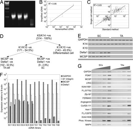
Considerable progress has been made in characterizing epidermal stem cells by microarray analysis of FACS-selected populations. One limitation of this approach is that the gene expression profiles represent the average of the cell population, potentially masking cellular heterogeneity of functional significance. To overcome this problem, we have performed single-cell expression profiling. We have generated cDNA libraries from single human epidermal cells, designated as stem or transit-amplifying cells on the basis of Delta1 and melanoma-associated chondroitin sulfate proteoglycan expression. Of the 14 putative stem cell markers identified, we selected one, the EGF receptor antagonist leucine-rich repeats and immunoglobulin-like domains 1 (Lrig1), for further study. Lrig1 was expressed in groups of basal cells in human interfollicular epidermis previously identified as enriched for stem cells. Overexpression of Lrig1 decreased keratinocyte proliferation but did not affect the proportion of stem and transit-amplifying cells, as judged by clonal growth characteristics. Down-regulation of Lrig1 using siRNA increased cell-surface EGF receptor levels, enhanced activation of downstream pathways, and stimulated proliferation. Lrig1 acted in part by negatively regulating the Myc promoter. We propose that Lrig1 maintains epidermal stem cells in a quiescent nondividing state, and that Lrig1 down-regulation triggers proliferation.


Conflict of interest statement

Conflict of interest statement: No conflicts declared.

Figures

Fig. 1.
Fig. 1.
Generation of single-cell cDNA libraries and marker validation. (A) Amplification of cDNA from 50 pg of total RNA from MCF7 cells. Triplicate samples are shown. (B) Real-time PCR demonstrating that relative expression levels of Gapdh, β1 Integrin, and Delta1 (in inverse order of abundance) compared with β-actin are preserved between amplified and nonamplified MCF7 cDNA samples. (C) Amplified, fragmented, and labeled cDNA from MCF7 and MCF10A cells was generated in hexaplicate and hybridized to HU133 Affymetrix gene arrays. The raw data were filtered as described in Materials and Methods. Fold values (y axis) for probes identified as differentially expressed using the single-cell method were truly differentially expressed when compared with fold values of the same samples analyzed using traditional methodology (x axis), because scatter is located in the upper-right and lower-left quadrants. (D) Markers used to identify cDNA libraries as originating from SC, TA cells, or terminally differentiated cells. Number of libraries of each type is shown in brackets. (E) Validation of marker expression by RT-PCR of cDNA libraries from individual SC and TA cells chosen for expression profiling. (F) Real-time PCR analysis of the picked cDNA libraries. (G) Validation of SC marker genes by semiquantitative PCR on total RNA from enriched populations of SC and TA cells by using a 4-fold dilution series of template.
Fig. 2.
Fig. 2.
Expression of Lrig1 in vivo and effects of overexpression in keratinocytes. (A–E) Immunofluorescence staining of human interfollicular epidermis with antibodies shown. (C–E) DAPI nuclear counterstain (blue). (A) Top (T) and bottom (B) of rete ridges are shown. The boxed area of C is shown at higher magnification in D. (Scale bars, 50 μm.) (F) Keratinocytes transduced with Lrig1 or empty retroviral vector (EV) were serum-starved for 24 h and stimulated with 10 ng/ml EGF for the number of minutes indicated. Western blots probed with antibodies are shown. Levels of P-ERK relative to tubulin were quantified by using NIH Image software, assigning the 0-min time point a value of 1. Data are representative of three independent experiments. (G) Percent BrdU-positive keratinocytes after transient transfection with eGFP or Lrig1. Mean ± SD of triplicate experiments (>100 cells per experiment). ∗P < 0.0001. (H) Flow cytometry of basal keratinocytes infected with empty retroviral vector (red) or Lrig1 (green). (I and J) Clonal growth assays of keratinocytes transduced with empty vector (EV) or Lrig1. (I) Representative dishes. (J) Mean ± SD of triplicate experiments.
Fig. 3.
Fig. 3.
Consequences of reducing Lrig1 expression by siRNA. (A) Relative levels of Lrig1 mRNA, determined by quantitative PCR, in keratinocytes transduced with Lrig1 siRNA or scrambled siRNA (scr). Values are expressed relative to 18S ribosomal RNA. Mean ± SD of quadruplicate experiments. ∗, P = 0.0004. (B) Western blots of keratinocytes transduced with Lrig1 siRNA or scrambled (scr) siRNA treated with EGF for the times indicated. Levels of P-ERK relative to tubulin controls were quantified using NIH Image software, assigning the 0-min time point a value of 1. Data are representative of three independent experiments. (C) Flow cytometry of basal keratinocytes transduced with scrambled siRNA (red) or Lrig1 siRNA (green). (D) Quantitation of cell number using Celltiter96. Data are means ± SD of hexaplicate samples. ∗, P < 0.0001. (E and F) Clonal growth assays. (E) Representative dishes. (F) Mean ± SD of triplicate samples. ∗, P = 0.0004. (G) Individual clones stained with anti-α6 integrin (green) and involucrin (red) antibodies. (Scale bar, 100 μm.)
Fig. 4.
Fig. 4.
Down-regulation of cMyc by Lrig1. (A) Western blot of keratinocytes transduced with Lrig1 or empty vector (EV) probed with antibodies to the proteins indicated. (B and C) Keratinocyte clone transduced with Lrig1 (green), stained for anti-cMyc (red) and DAPI (blue). (C) Higher magnification of boxed area in B. Cells with low Lrig1 and high Myc (arrows) or high Lrig1 and low Myc (arrowheads) are indicated. (Scale bars, 50 μm.) (D and E) Immunofluorescence staining of human epidermis with antibodies shown and DAPI nuclear counterstain (blue). Confocal images (Z stacks) of serial sections are shown. (Scale bar, 25 μm.) (F) Activity of Myc promoter (P1P2) determined by dual luciferase assays, showing percent induction by EGF in keratinocytes transduced with Lrig1 or empty vector (EV). Mean ± SD of quadruplicate experiments. ∗, P = 0.002. (G) Myc promoter activity in keratinocytes stimulated with EGF or keratinocyte serum-free medium in the presence or absence of AG1478. Mean ± SD of triplicate experiments. ∗∗, P = 0.0002; ∗∗∗, P < 0.0001. (H) Cells transduced with MycER or EV were transiently transfected with Lrig1 or GFP and either treated with 4OHT or untreated. Percent transfected cells that incorporated BrdU during a 24-h label is shown. Data represent mean ± SD of four or more samples with >100 transfected cells per sample. (∗∗∗, P < 0.0001; ∗∗, P = 0.0002). (I) Model for the role of Lrig1 in maintaining SC in a quiescent nondividing state.

References

    1. Owens D. M., Watt F. M. Nat. Rev. Cancer. 2003;3:444–451. - PubMed
    1. Alonso L., Fuchs E. Proc. Natl. Acad. Sci. USA. 2003;100(Suppl. 1):11830–11835. - PMC - PubMed
    1. Tumbar T., Guasch G., Greco V., Blanpain C., Lowry W. E., Rendl M., Fuchs E. Science. 2004;303:359–363. - PMC - PubMed
    1. Morris R. J., Liu Y., Marles L., Yang Z., Trempus C., Li S., Lin J. S., Sawicki J. A., Cotsarelis G. Nat. Biotechnol. 2004;22:411–417. - PubMed
    1. Cotsarelis G., Sun T. T., Lavker R. M. Cell. 1990;61:1329–1337. - PubMed

Publication types

Substances

Associated data