. 2006 Jul 28;34(13):e93.
doi: 10.1093/nar/gkl515.

Real-time detection and continuous monitoring of ER stress in vitro and in vivo by ES-TRAP: evidence for systemic, transient ER stress during endotoxemia

Affiliations

Real-time detection and continuous monitoring of ER stress in vitro and in vivo by ES-TRAP: evidence for systemic, transient ER stress during endotoxemia

Nobuhiko Hiramatsu et al. Nucleic Acids Res. .

Abstract

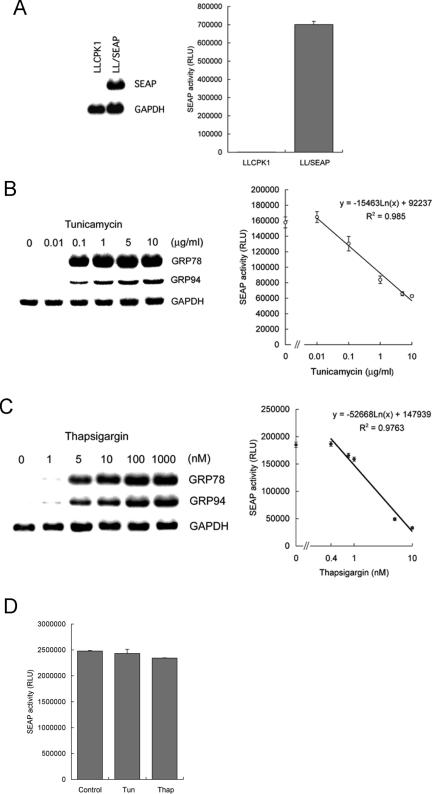
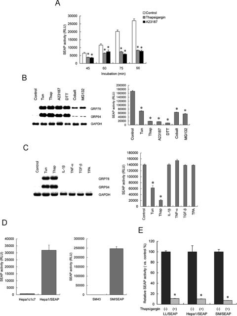
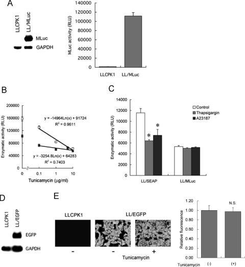
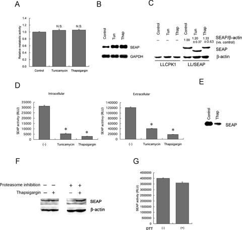
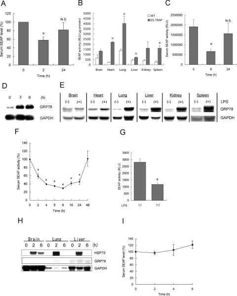
Activity of secreted alkaline phosphatase (SEAP) produced by transfected cells is rapidly down-regulated by endoplasmic reticulum (ER) stress independent of transcriptional regulation. This phenomenon was observed in a wide range of cell types triggered by various ER stress inducers. The magnitude of the decrease in SEAP was proportional to the extent of ER stress and inversely correlated with the induction of endogenous ER stress markers grp78 and grp94. In contrast to SEAP, activity of secreted luciferase was less susceptible to ER stress. The decrease in SEAP activity by ER stress was caused by abnormal post-translational modification, accelerated degradation and reduced secretion of SEAP protein. In transgenic mice constitutively producing SEAP, systemic induction of ER stress led to reduction in serum SEAP. In these mice, administration with lipopolysaccharide caused rapid, transient decrease in serum SEAP activity, and it was correlated with up-regulation of grp78 in several organs including the spleen, lung, kidney, liver and heart. These results elucidated for the first time a possible involvement of transient, systemic ER stress in endotoxemia and provided evidence for usefulness of ER stress responsive alkaline phosphatase for real-time monitoring of ER stress in vitro and in vivo.


Figures

Figure 1
Figure 1
Suppression of SEAP activity by endoplasmic reticulum (ER) stress in stably transfected cells. (A) Establishment of stable transfectants. LLCPK1 cells were stably transfected with a SEAP gene under the control of the simian virus (SV40) promoter, and established LL/SEAP cells were subjected to northern blot analysis (left) and chemiluminescent assay of culture medium (right). (B and C) Responses of LL/SEAP cells to tunicamycin (B) and thapsigargin (C). LL/SEAP cells were treated with tunicamycin (0.01–10 µg/ml) or thapsigargin (0.4–1000 nM) for 6 h, and expression of grp78 and grp94 (left) and activity of SEAP in culture media (right) were evaluated. (D) Recombinant SEAP was added with tunicamycin (Tun, 10 µg/ml) or thapsigargin (Thap, 1 µM) and incubation at 37°C for 6 h. Activity of SEAP was assessed by chemiluminescent assay. SEAP assays were performed in quadruplicate, and data are expressed as means ± SE. RLU, relative light unit. In northern blot analysis, expression of GAPDH was used as a loading control.
Figure 2
Figure 2
Characterization of the response of SEAP to ER stress. (A) Rapid reduction in SEAP activity by ER stress. LL/SEAP cells were pretreated with thapsigargin (1 µM) or A23187 (2 µM) for 30 min. After changing the medium, the cells were further incubated in the presence of the drugs for up to 60 min. Culture media were collected every 15 min and subjected to SEAP assay. (B) Responses to known ER stress inducers. LL/SEAP cells were treated with tunicamycin (Tun; 10 µg/ml), thapsigargin (Thap; 1 µM), A23187 (2 µM), dithiothreitol (DTT; 1 mM), cobalt chloride (Cobalt; 1 mM) or MG132 (50 µM) for 6 h and subjected to northern blot analysis of grps (left) and SEAP assay (right). (C) Lack of responses to ER stress-unrelated stimuli. LL/SEAP cells were treated with interleukin 1β (IL-1β; 20 ng/ml), tumor necrosis factor-α (TNF-α; 250 U/ml), transforming growth factor-β (TGF-β; 10 ng/ml) or 12-o-tetradecanoylphorbol-13-acetate (TPA; 50 ng/ml) for 6 h and subjected to analyses. Tunicamycin (10 µg/ml) and thapsigargin (1 µM) were used as positive controls. (D) Secretion of SEAP by Hepa1/SEAP and SM/SEAP cells. The murine hepatoma cell line Hepa-1c1c7 and the rat mesangial cell line SM43 were stably transfected with a SEAP gene under the control of the SV40 promoter, and Hepa1/SEAP and SM/SEAP cells were established. Activity of SEAP in culture media was evaluated by chemiluminescent assay. (E) Responses to ER stress of LL/SEAP and Hepa1/SEAP cells. LL/SEAP, Hepa1/SEAP and SM/SEAP cells were treated with (+) or without (−) thapsigargin (1 µM) for 6 h, and culture media were subjected to SEAP assay. Assays were performed in quadruplicate, and data are expressed as means ± SE. Asterisks indicate statistically significant differences (P < 0.05).
Figure 3
Figure 3
Responses of other secreted and non-secreted reporter proteins to ER stress. (A) Establishment of LL/MLuc cells. LLCPK1 cells were stably transfected with a gene encoding secreted Metridia luciferase (MLuc), and LL/MLuc cells were established. left, northern blot analysis of MLuc mRNA in LLCPK1 cells and LL/MLuc cells. right, MLuc activity in culture media of LLCPK1 cells and LL/MLuc cells. (B) Dose-dependent responses of MLuc and SEAP to tunicamycin. LL/MLuc cells (closed square) and LL/SEAP cells (open square) were treated with tunicamycin (0.1–10 µg/ml) for 6 h, and culture media were subjected to chemiluminescent assays. (C) Susceptibility of MLuc and SEAP to ER stress triggered by thapsigargin and A23187. LL/SEAP and LL/MLuc cells were pretreated with thapsigargin (1 µM) or A23187 (2 µM) for 30 min. After changing the medium, the cells were further incubated in the presence of the drugs for 30 min and subjected to chemiluminescent assays. (D) Establishment of LL/EGFP cells. LLCPK1 cells were stably transfected with a gene encoding enhanced green fluorescent protein (EGFP), and LL/EGFP cells were established. Northern blot analysis of EGFP mRNA in LLCPK1 cells and LL/EGFP cells is shown. (E) Effect of ER stress on the activity of EGFP. Fluorescence microscopic analysis of LLCPK1 cells and LL/EGFP cells was performed before (−) and after (+) the treatment with tunicamycin (10 µg/ml) for 8 h. Fluorescence intensity of EGFP was evaluated, and relative fluorescence was shown on the right. All assays were performed in quadruplicate, and data are expressed as means ± SE. Asterisks indicate statistically significant differences (P < 0.05). N.S., not significant.
Figure 4
Figure 4
Mechanisms involved in the suppression of SEAP activity by ER stress. (AE) LL/SEAP cells were treated with tunicamycin (10 µg/ml) or thapsigargin (1 µM) for 6 h and subjected to analyses. (A) Cell viability evaluated by formazan assay. (B) Northern blot analysis of SEAP mRNA. (C) Western blot analysis of intracellular levels of SEAP. As a loading control, the level of β-actin was examined (bottom), and relative values of SEAP against β-actin (SEAP/β-actin) were shown (n = 3). (D) Activity of intracellular (left) and extracellular (right) SEAP. (E) Western blot analysis of extracellular levels of SEAP. (F) Intracellular levels of SEAP in thapsigargin-treated cells with or without blockade of the proteasome pathway. LL/SEAP cells were treated with thapsigargin in the absence (−) or presence (+) of proteasome inhibition (25 µM MG132) for 12 h, and cellular protein was subjected to western blot analysis. (G) Lack of involvement of disulfide bonds formation in SEAP activation. SEAP protein was treated with (+) or without (−) 100 mM DTT for 1 h. DTT was washed out using cut-off membranes (M.W. < 50 kDa), and the resultant samples were subjected to SEAP assay. All SEAP assays were performed in quadruplicate, and data are expressed as means ± SE. Asterisks indicate statistically significant differences (P < 0.05). N.S., not significant.
Figure 5
Figure 5
Detection and real-time monitoring of ER stress during endotoxemia using ES-TRAP. (A) Effect of systemic ER stress on the level of serum SEAP in ex vivo gene transferred mice. Hepa1/SEAP cells (2.5 × 10⁵ cells) were implanted into the peritoneal cavity of C57BL/6 mice. After 24–48 h, thapsigargin (1 mg/kg body weight) was administered into eight mice. Serum was sampled at 2 and 24 h, and activity of SEAP was evaluated. (B) Activity of SEAP in organs of transgenic ES-TRAP mice. Tissue extracts were prepared from perfused organs of wild-type mice (WT) and ES-TRAP mice and subjected to chemiluminescent assay, as described in Materials and Methods. Activity of SEAP per 1 µg total protein was evaluated and compared among different organs. (C) Effect of systemic ER stress on the level of serum SEAP in ES-TRAP mice. Thapsigargin (1 mg/kg body weight) was administered into four mice (body weight 25 g) intraperitoneally, and serum was sampled at 8 and 24 h. (D) Induction of ER stress by LPS in macrophages. NR8383 alveolar macrophages were exposed to LPS (1 µg/ml) for 3–6 h, and expression of grp78 was examined by northern blot analysis. (EG) Effects of endotoxemia in ES-TRAP mice on induction of ER stress in various organs (E), serum SEAP activity (F) and tissue SEAP activity in the spleen (G). ES-TRAP mice (body weight 20 g, n = 8) were administered without (−) or with (+) LPS (200 µg/mouse) intraperitoneally, and organs (8 h) and serum (2–48 h) were collected for northern blot analysis of grp78 (E) and SEAP assay (F and G). In (G), activity of SEAP in the spleen before [LPS(−)] and 8 h after the administration with LPS [LPS(+)] is shown. (H and I) Lack of in vivo response of SEAP to hyperthermia. ES-TRAP mice (body weight 22 g, n = 4) were heated at 42°C for 15 min, and brains, lungs and livers were collected before or 2 and 6 h after the whole body hyperthermia. Expression of hsp70 and grp78 was examined by northern blot analysis (H). Serum was also sampled every 2 h until 6 h and subjected to chemiluminescent assay to evaluate activity of SEAP (I). Data are expressed as means ± SE. Asterisks indicate statistically significant differences (P < 0.05). N.S., not significant.

Similar articles

Cited by

References

    1. Aridor M., Balch W.E. Integration of endoplasmic reticulum signaling in health and disease. Nature Med. 1999;5:745–751. - PubMed
    1. Schroder M., Kaufman R.J. ER stress and the unfolded protein response. Mutat. Res. 2005;569:29–63. - PubMed
    1. Kozutsumi Y., Segal M., Normington K., Gething M.J., Sambrook J. The presence of malfolded proteins in the endoplasmic reticulum signals the induction of glucose-regulated proteins. Nature. 1988;332:462–464. - PubMed
    1. Nakagawa T., Zhu H., Morishima N., Li E., Xu J., Yankner B.A., Yuan J. Caspase-12 mediates endoplasmic reticulum-specific apoptosis and cytotoxicity by amyloid-β. Nature. 2003;403:98–103. - PubMed
    1. He H., McColl K., Distelhorst C.W. Involvement of c-Fos in signaling grp78 induction following ER calcium release. Oncogene. 2000;19:5936–5943. - PubMed

Publication types

MeSH terms