. 2006;2006(1):32713.
doi: 10.1155/JBB/2006/32713.

The potential regulation of L1 mobility by RNA interference

Affiliations

The potential regulation of L1 mobility by RNA interference

Shane R Horman et al. J Biomed Biotechnol. 2006.

Abstract

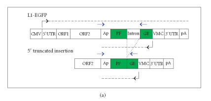
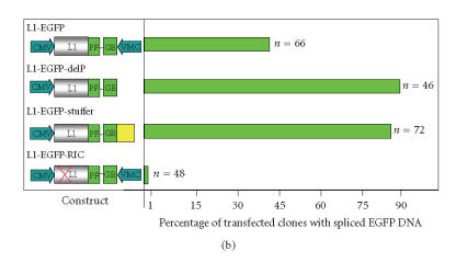
The hypothesis that RNA interference constrains L1 mobility seems inherently reasonable: L1 mobility can be dangerous and L1 RNA, the presumed target of RNAi, serves as a critical retrotransposition intermediate. Despite its plausibility, proof for this hypothesis has been difficult to obtain. Studies attempting to link the L1 retrotransposition frequency to alterations in RNAi activity have been hampered by the long times required to measure retrotransposition frequency, the pleiotropic and toxic effects of altering RNAi over similar time periods, and the possibility that other cellular machinery may contribute to the regulation of L1s. Another problem is that the commonly used L1 reporter cassette may serve as a substrate for RNAi. Here we review the L1-RNAi hypothesis and describe a genetic assay with a modified reporter cassette that detects approximately 4 times more L1 insertions than the conventional retrotransposition assay.


Figures

Figure 1
Figure 1
Generation of L1 dsRNA. L1 dsRNA could arise from different transcripts (shown in Figures 1(a), 1(b), and 1(c)) or from the same transcript (shown in Figure 1(d)). (a) An L1 transcript originates from the internal 5′UTR promoter, producing a sense-strand RNA. Neighboring the same element, an antisense-oriented heterologous promoter produces a read-through transcript that includes antisense L1 RNA. (b) An L1 is transcribed producing sense RNA and another L1 insertion, elsewhere in the genome, is transcribed off of a heterologous promoter yielding an antisense RNA. (c) An L1 is transcribed off of its 5′UTR producing a sense transcript, while antisense promoter activity of the 5′UTR produces an antisense transcript. (d) If an L1 inserts near another L1 sequence in the genome, it may be possible to create a hairpin. The figure shows two full-length L1 sequences facing each other, although it should be noted that hairpins could also form between truncated L1 copies that face each other, as long as there is a transcript that extends between the copies. DNA strands are shown with solid lines and RNA with dashed lines. In the scenario depicted, transcripts off of either DNA strand extending through the two L1 sequences will give rise to self-complementary regions: the forward facing L1 and the reverse complementary sequence of L1 on the same RNA strand can base pair, forming hairpins (dashed vertical lines) with stretches of dsRNA.
Figure 1
Figure 1
Generation of L1 dsRNA. L1 dsRNA could arise from different transcripts (shown in Figures 1(a), 1(b), and 1(c)) or from the same transcript (shown in Figure 1(d)). (a) An L1 transcript originates from the internal 5′UTR promoter, producing a sense-strand RNA. Neighboring the same element, an antisense-oriented heterologous promoter produces a read-through transcript that includes antisense L1 RNA. (b) An L1 is transcribed producing sense RNA and another L1 insertion, elsewhere in the genome, is transcribed off of a heterologous promoter yielding an antisense RNA. (c) An L1 is transcribed off of its 5′UTR producing a sense transcript, while antisense promoter activity of the 5′UTR produces an antisense transcript. (d) If an L1 inserts near another L1 sequence in the genome, it may be possible to create a hairpin. The figure shows two full-length L1 sequences facing each other, although it should be noted that hairpins could also form between truncated L1 copies that face each other, as long as there is a transcript that extends between the copies. DNA strands are shown with solid lines and RNA with dashed lines. In the scenario depicted, transcripts off of either DNA strand extending through the two L1 sequences will give rise to self-complementary regions: the forward facing L1 and the reverse complementary sequence of L1 on the same RNA strand can base pair, forming hairpins (dashed vertical lines) with stretches of dsRNA.
Figure 2
Figure 2
(a) The standard L1 reporter construct contains opposing promoters. The standard L1 reporter construct used in our laboratory (L1-EGFP) consists of the CMV promoter, the human L1RP element, and the antisense EGFP gene cloned into the L1 3′UTR followed by the SV40 late poly-A sequence in pCEP4 (construct described in more detail in [51]). When L1-EGFP retrotransposes, a full-length L1 RNA is transcribed, the intron interrupting EGFP is spliced out, and the processed RNA is reverse transcribed, and a cDNA copy is inserted into the genome. If the insertion is of sufficient length and enters the genome in a transcriptionally permissive region, retrotransposition can be detected phenotypically by screening for EGFP expression. Retrotransposition can also be assayed genetically by performing PCR with primers that flank the EGFP intron. Because the EGFP marker is driven off of an antisense-oriented promoter relative to the L1, the potential exists for creating dsRNA. L1 and EGFP transcripts are given by dashed horizontal lines, promoters are denoted with black arrows, and blue arrows indicate intron-flanking primers used to distinguish new insertions from the parental L1. (b) Loss of an antisense promoter increases L1 retrotransposition in a cultured cell assay. 143B osteosarcoma cells were transfected with one of the following constructs as shown in Figure 2(a): L1-EGFP (the same wild-type L1 retrotransposition construct shown in Figure 2(a)), L1-EGFP-DelP (identical to L1-EGFP except that the CMV promoter driving EGFP was deleted), L1-EGFP-Stuffer (identical to L1-EGFP except that the CMV promoter driving EGFP was replaced with a piece of DNA lacking promoter activity or polyadenylation signals), or L1-EGFP-RIC (retrotransposition incompetent due to two missense mutations (marked with a red X over the L1 coding sequence) derived from the JM111 L1 mutant [52]). Boxes indicate coding sequences except for the yellow box in the L1-EGFP-Stuffer construct that denotes the stuffer sequence. Arrows denote the promoters and the black line separating the EGFP cassette denotes the intron. Cells were selected in hygromycin for two weeks and individual clones were picked and expanded. PCR using primers that flank the intron/exon splice site in EGFP (as described in [11]) was used to monitor individual clones of antibiotic-resistant cells for L1 retrotransposition (loss of the intron in EGFP). The percentages of clones that had the spliced EGFP are shown in Figure 2(b). The number of clones surveyed for each genotype is given to the right of each of the bars. None of the retrotransposition incompetent L1 transfectants had a spliced EGFP product.
Figure 2
Figure 2
(a) The standard L1 reporter construct contains opposing promoters. The standard L1 reporter construct used in our laboratory (L1-EGFP) consists of the CMV promoter, the human L1RP element, and the antisense EGFP gene cloned into the L1 3′UTR followed by the SV40 late poly-A sequence in pCEP4 (construct described in more detail in [51]). When L1-EGFP retrotransposes, a full-length L1 RNA is transcribed, the intron interrupting EGFP is spliced out, and the processed RNA is reverse transcribed, and a cDNA copy is inserted into the genome. If the insertion is of sufficient length and enters the genome in a transcriptionally permissive region, retrotransposition can be detected phenotypically by screening for EGFP expression. Retrotransposition can also be assayed genetically by performing PCR with primers that flank the EGFP intron. Because the EGFP marker is driven off of an antisense-oriented promoter relative to the L1, the potential exists for creating dsRNA. L1 and EGFP transcripts are given by dashed horizontal lines, promoters are denoted with black arrows, and blue arrows indicate intron-flanking primers used to distinguish new insertions from the parental L1. (b) Loss of an antisense promoter increases L1 retrotransposition in a cultured cell assay. 143B osteosarcoma cells were transfected with one of the following constructs as shown in Figure 2(a): L1-EGFP (the same wild-type L1 retrotransposition construct shown in Figure 2(a)), L1-EGFP-DelP (identical to L1-EGFP except that the CMV promoter driving EGFP was deleted), L1-EGFP-Stuffer (identical to L1-EGFP except that the CMV promoter driving EGFP was replaced with a piece of DNA lacking promoter activity or polyadenylation signals), or L1-EGFP-RIC (retrotransposition incompetent due to two missense mutations (marked with a red X over the L1 coding sequence) derived from the JM111 L1 mutant [52]). Boxes indicate coding sequences except for the yellow box in the L1-EGFP-Stuffer construct that denotes the stuffer sequence. Arrows denote the promoters and the black line separating the EGFP cassette denotes the intron. Cells were selected in hygromycin for two weeks and individual clones were picked and expanded. PCR using primers that flank the intron/exon splice site in EGFP (as described in [11]) was used to monitor individual clones of antibiotic-resistant cells for L1 retrotransposition (loss of the intron in EGFP). The percentages of clones that had the spliced EGFP are shown in Figure 2(b). The number of clones surveyed for each genotype is given to the right of each of the bars. None of the retrotransposition incompetent L1 transfectants had a spliced EGFP product.

Similar articles

Cited by

References

    1. Tomari Y, Zamore PD. Perspective: machines for RNAi. Genes & Development. 2005;19(5):517–529. - PubMed
    1. Carmell MA, Hannon GJ. RNase III enzymes and the initiation of gene silencing. Nature Structural & Molecular Biology. 2004;11(3):214–218. - PubMed
    1. Plasterk RHA. RNA silencing: the genome's immune system. Science. 2002;296(5571):1263–1265. - PubMed
    1. Bagasra O, Prilliman KR. RNA interference: the molecular immune system. Journal of Molecular Histology. 2004;35(6):545–553. - PubMed
    1. Sijen T, Plasterk RHA. Transposon silencing in the Caenorhabditis elegans germ line by natural RNAi. Nature. 2003;426(6964):310–314. - PubMed

LinkOut - more resources