Comparative Study
. 2006 Aug;116(8):2152-60.
doi: 10.1172/JCI28084. Epub 2006 Jul 27.

Glucocorticoids suppress bone formation via the osteoclast

Affiliations
Comparative Study

Glucocorticoids suppress bone formation via the osteoclast

Hyun-Ju Kim et al. J Clin Invest. 2006 Aug.

Abstract

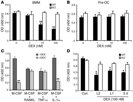
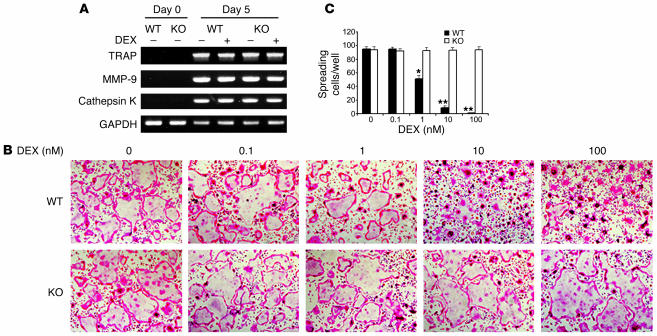
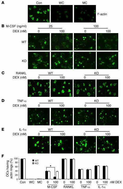
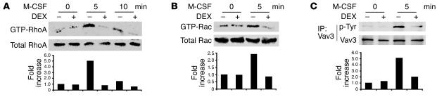
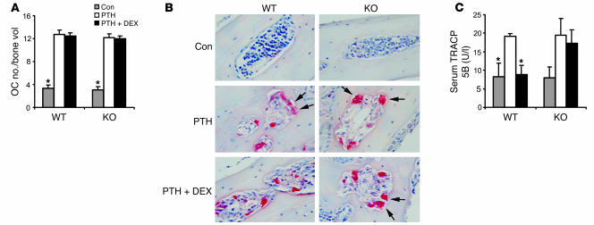
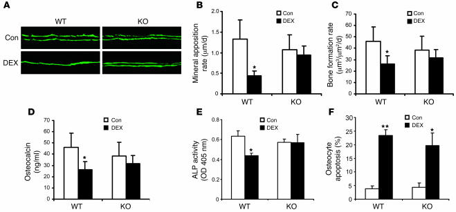
The pathogenesis of glucocorticoid-induced (GC-induced) bone loss is unclear. For example, osteoblast apoptosis is enhanced by GCs in vivo, but they stimulate bone formation in vitro. This conundrum suggests that an intermediary cell transmits a component of the bone-suppressive effects of GCs to osteoblasts in the intact animal. Bone remodeling is characterized by tethering of the activities of osteoclasts and osteoblasts. Hence, the osteoclast is a potential modulator of the effect of GCs on osteoblasts. To define the direct impact of GCs on bone-resorptive cells, we compared the effects of dexamethasone (DEX) on WT osteoclasts with those derived from mice with disruption of the GC receptor in osteoclast lineage cells (GRoc-/- mice). While the steroid prolonged longevity of osteoclasts, their bone-degrading capacity was suppressed. The inhibitory effect of DEX on bone resorption reflects failure of osteoclasts to organize their cytoskeleton in response to M-CSF. DEX specifically arrested M-CSF activation of RhoA, Rac, and Vav3, each of which regulate the osteoclast cytoskeleton. In all circumstances GRoc-/- mice were spared the impact of DEX on osteoclasts and their precursors. Consistent with osteoclasts modulating the osteoblast-suppressive effect of DEX, GRoc-/- mice are protected from the steroid's inhibition of bone formation.


Figures

Figure 1
Figure 1. DEX impacts osteoclast lineage cells in a differentiation- and cytokine-dependent manner.
WT and GRoc–/– BMMs were cultured with M-CSF (50 ng/ml) in the absence (A) or presence (B) of RANKL (30 ng/ml) with various concentrations of DEX. After 3 days, proliferation was assessed by BrdU incorporation. (C) WT BMMs were incubated with or without DEX (100 nM) in the presence of M-CSF alone or with RANKL, TNF-α (10 ng/m) or IL-1α (10 ng/ml) for 3 days. Proliferation was evaluated by BrdU incorporation assay. (D) WT and GRoc–/– BMMs were cultured with M-CSF and RANKL for 5 days to generate mature osteoclasts. DEX (100 nM) or vehicle was added for the entire 5-day culture period or the last 2 days (L2) or 1 day (L1). After 5 days, the relative apoptosis rate was determined by ELISA. *P < 0.01, **P < 0.001 within each group.
Figure 2
Figure 2. DEX-treated BMMs differentiate into osteoclasts but fail to spread.
(A) WT and GRoc–/– BMMs were cultured with M-CSF and RANKL with or without DEX (100 nM). At day 0 and day 5, RNA was extracted, and the expression of osteoclastogenic markers was analyzed by RT-PCR. GAPDH served as loading control. (B) WT and GRoc–/– BMMs were cultured with M-CSF and RANKL with or without increasing concentrations of DEX. Five-day osteoclastogenic cultures were stained for TRAP activity. Magnification, ×250. (C) Statistical analysis of the number of WT and GRoc–/– spread TRAP-positive multinucleated cells/well. *P < 0.001, **P < 0.0001 versus DEX-untreated WT.
Figure 3
Figure 3. DEX inhibits spreading in the later stage of osteoclast differentiation.
WT and GRoc–/– BMMs were cultured for 5 days in M-CSF and RANKL and stained for TRAP activity. The cells were treated with DEX (100 nM) for the entire 5 days, the first 3 days (F3), the first 4 days, the last 2 days, or the last day. Control cultures were maintained in the absence of DEX for 5 days. (A) Duration of exposure to Dex in days. (B) Osteoclastogenic cultures stained for TRAP activity. Magnification, ×250.
Figure 4
Figure 4. DEX inhibits M-CSF–induced actin ring formation.
WT and GRoc–/– BMMs were plated on dentin and cultured with M-CSF (10 ng/ml) and RANKL (100 ng/ml). (A) After 5 days, WT cells were fixed before (Con), after washing with cold PBS (washing control [WC]), or after washing and incubation in α-MEM/10% FBS (media control [MC]) for 5 hours. The fixed cells were stained with FITC-phalloidin to visualize actin rings. (BE) After 5 days WT and GRoc–/– cells were washed with cold PBS and incubated with or without DEX (100 nM) in the presence of M-CSF (25 or 100 ng/ml) (B), RANKL (100 ng/ml) (C), TNF-α (10 ng/ml) (D), or IL-1α (10 ng/ml) (E). After 5 hours incubation, the fixed cells were stained with FITC-phalloidin. (F) The percentage of osteoclasts (OCs) with recovered actin ring formation with or without DEX and various cytokines. *P < 0.001. Magnification, ×250.
Figure 5
Figure 5. DEX inhibits M-CSF–induced RhoA, Rac, and Vav3 activation.
WT BMMs maintained in M-CSF and RANKL for 3 days were exposed to DEX (100 nM) or vehicle for 16 hours and then stimulated with M-CSF (100 ng/ml). GTP-RhoA and GTP-Rac were isolated by GST pull-down and immunoblotted with RhoA- (A) or Rac-specific (B) antibodies. (C) Vav3 was immunoprecipitated from the cell lysate and phosphotyrosine (p-Tyr) content of the immunoprecipitate determined by immunoblot with anti-phosphotyrosine antibody. In all circumstances, the data are expressed as fold difference in DEX-exposed (+) relative to vehicle-exposed (–) cells.
Figure 6
Figure 6. DEX suppresses osteoclastic bone resorption in vitro.
(A) WT and GRoc–/– BMMs were cultured on dentin with RANKL and M-CSF with or without DEX. After 5 days, resorption pits were stained with toluidine blue (arrows). (B) WT and GRoc–/– BMMs were cultured on plastic in the presence of M-CSF and RANKL. After 3 days the cells were lifted and replated on dentin in M-CSF and RANKL with or without DEX. Three days later, resorption pits were stained with toluidine blue. Magnification, ×100.
Figure 7
Figure 7. DEX suppresses PTH-stimulated bone resorption in vivo.
WT and GRoc–/– mice were injected daily for 4 days with vehicle (Con) or PTH or PTH plus DEX (n = 4). Histological sections of calvariae were stained for TRAP activity. (A) Osteoclast number/unit bone volume. (B) TRAP-stained histological sections of calvariae (arrows indicate osteoclasts with ruffled membranes in resorption lacunae). Magnification, ×250. (C) Serum TRACP5b levels at sacrifice. *P < 0.01 versus PTH-treated WT.
Figure 8
Figure 8. GRoc–/– mice are protected from DEX-suppressed bone formation.
WT and GRoc–/– mice were injected with DEX (10 mg/kg) or vehicle daily for 14 days. Calcein was administered 8 and 2 days prior to sacrifice. (A) Fluorescent micrographs of representative calcein labels. Magnification, ×100. (B) Measured mineral apposition rate. (C) Measured bone formation rate. (D) Serum osteocalcin levels at sacrifice. (E) Serum alkaline phosphatase (ALP) levels at sacrifice. (F) Percentage of osteocytes in histological sections of bone undergoing apoptosis. *P < 0.05, **P < 0.005 versus control.

References

    1. Weinstein R.S. Glucocorticoid-induced osteoporosis. Rev. Endocr. Metab. Disord. 2001;2:65–73. - PubMed
    1. Weinstein R.S., Jilka R.L., Parfitt A.M., Manolagas S.C. Inhibition of osteoblastogenesis and promotion of apoptosis of osteoblasts and osteocytes by glucocorticoids. J. Clin. Invest. 1998;102:274–282. - PMC - PubMed
    1. Pereira R.M., Delany A.M., Canalis E. Cortisol inhibits the differentiation and apoptosis of osteoblasts in culture. Bone. 2001;28:484–490. - PubMed
    1. Pereira R.C., Delany A.M., Canalis E. Effects of cortisol and bone morphogenetic protein-2 on stromal cell differentiation: correlation with CCAAT-enhancer binding protein expression. Bone. 2002;30:685–691. - PubMed
    1. Aubin J.E. Osteoprogenitor cell frequency in rat bone marrow stromal populations: role for heterotypic cell-cell interactions in osteoblast differentiation. J. Cell. Biochem. 1999;72:396–410. - PubMed

Publication types

MeSH terms