Case Reports
. 2006;33(2):171-9.

Symptomatic anomalous origination of the left coronary artery from the opposite sinus of valsalva. Clinical presentations, diagnosis, and surgical repair

Affiliations
Case Reports

Symptomatic anomalous origination of the left coronary artery from the opposite sinus of valsalva. Clinical presentations, diagnosis, and surgical repair

Paolo Angelini et al. Tex Heart Inst J. 2006.

Abstract

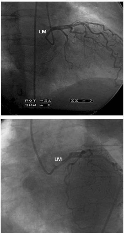
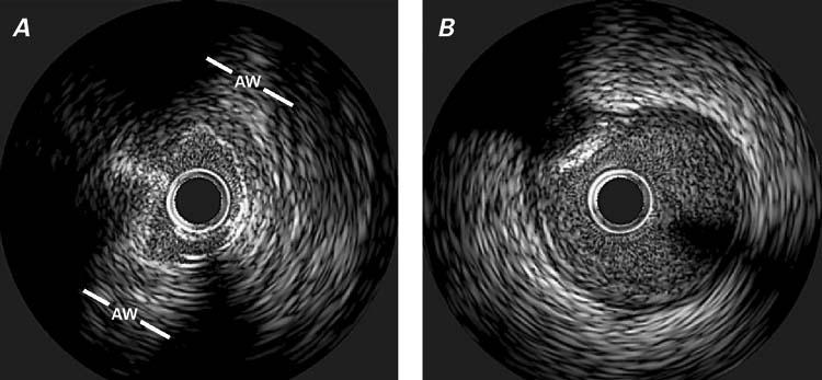
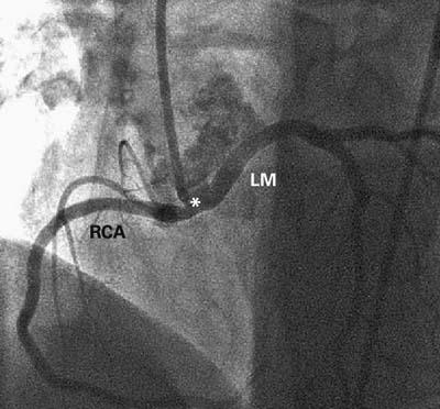
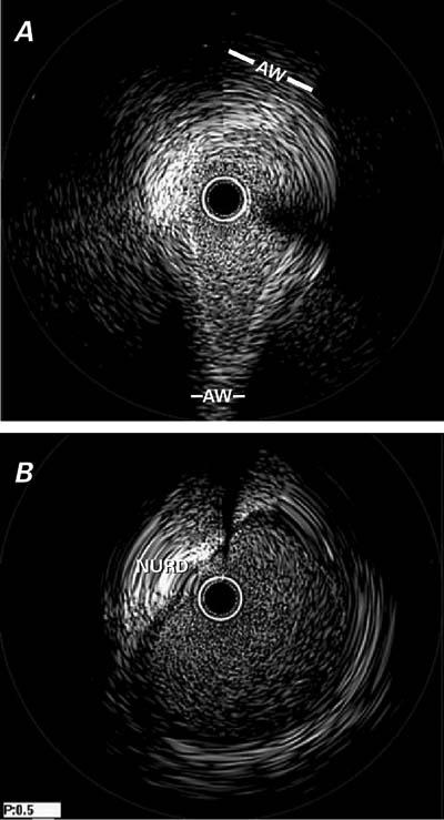
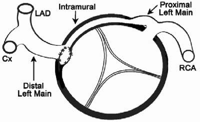
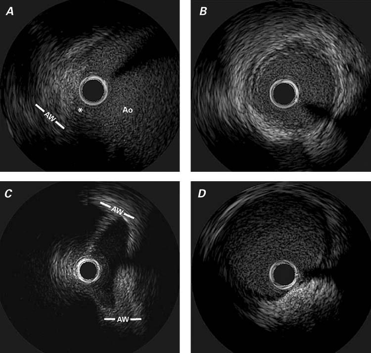
Anomalous origination of a coronary artery can have serious, even fatal, consequences. Intravascular ultrasonography has recently provided new insights into anomalous coronary artery origination from the opposite sinus of Valsalva. On the basis of these insights, we describe 3 typical forms of this anomaly with left coronary artery involvement, including clinical presentations, diagnostic methods (particularly intravascular ultrasonography), and details of surgical treatment. In this case series, the left coronary artery originated from the noncoronary sinus in 1 patient and from the right sinus in another patient. In the 3rd patient, both the left and right coronary arteries originated from the ascending aorta above the sinotubular junction. Baseline areas of stenosis ranged from 48.6% to 70.1%. Intravascular ultrasonography was the only method that enabled us to clarify the mechanisms and the severity of the anomaly. Pharmacologic challenge was useful to predict worsening that might have occurred under physiologic conditions. We found that, in cases of symptomatic left anomalous coronary artery origination from the opposite sinus of Valsalva, the proximal segment of the left coronary artery consistently has (1) an intramural course inside the aortic wall; (2) hypoplasia, as determined by its circumference; and (3) a cross-sectional ovaloid deformity (lateral compression) with phasic and exercise-induced worsening of the deformity With regard to surgical treatment, ostioplasty is preferable to coronary bypass. To establish sound guidelines for managing these anomalies, a larger series should be studied prospectively with quantitative parameters and long-term follow-up.


Figures

None
Fig. 1 Case 1. Selective left coronary angiogram in the right anterior oblique (A) and left anterior oblique projections (B). The left main trunk is unusually long and originates quite posteriorly. Subselective injections provided clear evidence that the ostium was located just to the right of the posterior commissure, at the noncoronary sinus (not shown). In (A), it is apparent that the proximal left coronary artery is located at the posterior border of the aortic root, which corresponds to the posterior commissure, in the right anterior oblique projection. LM = left main
None
Fig. 2 Case 1. Intravascular ultrasonographic images of the left main trunk at the intramural (aortic) segment (A) and the more distal (epicardial) segment (B). In (A), note the reduced lumen of the proximal segment in the absence of an atherosclerotic intimal build-up, which is present in the distal segment (in [B] at 10 o'clock there is a calcific plaque). AW = aortic wall
None
Fig. 3 Case 2. Selective angiogram of the common coronary artery in the left anterior oblique cranial projection. The common trunk (*) is short (about 10 mm) and arises orthogonally from the aortic wall before bifurcating into the right coronary artery (RCA), which has a normal course, and the anomalous left main artery (LM), which wraps around the aortic root.
None
Fig. 4 Case 2. Intravascular ultrasonographic images of the intramural (A) and extramural (B) segments of the left main trunk. In (A), note the severe hypoplasia in the proximal segment (which is not apparent on the angiogram in Fig. 3), the absence of intimal thickening, and the ovaloid cross-section; in contrast, the distal left main artery has a round lumen and an intimal plaque (B). See text and Table I for further details. AW = aortic wall; NURD = non-uniform rotational distortion
None
Fig. 5 Case 2. Diagram of the surgical repair shows a cross-sectional view of the aortic root. The new, surgically-created ostium provides unrestricted direct access to the extramural portion of the left main artery, which is distal to the intramural hypoplastic proximal segment. Cx = circumflex artery; LAD = left anterior descending coronary artery; RCA = right coronary artery
None
Fig. 6 Case 3. A) Angiogram of the right coronary artery (RCA) in the left anterior oblique projection. Overspilling of the contrast agent identifies the adjacent left coronary ostium (LCA) and the left aortic sinus of Valsalva (LSV). There is no evidence of ostial stenosis. B) Angiogram of the left coronary artery, in the posteroanterior projection, suggests the presence of a membrane-like ostial stenosis.
None
Fig. 7 Case 3. Intravascular ultrasonographic images of the proximal right coronary artery (ostial, A and distal reference, B) segments and the left (C, D) coronary artery. Figure C was obtained during forceful injection of saline (which led to the enhancement of luminal definition). See Table I for related area measurements. * = ostium; Ao = aortic lumen; AW = aortic wall

Similar articles

Cited by

References

    1. Angelini P, Velasco JA, Flamm S. Coronary anomalies: incidence, pathophysiology, and clinical relevance. Circulation 2002;105:2449–54. - PubMed
    1. Hariharan R, Kacere RD, Angelini P. Can stent-angioplasty be a valid alternative to surgery when revascularization is indicated for anomalous origination of a coronary artery from the opposite sinus? Tex Heart Inst J 2002;29:308–13. - PMC - PubMed
    1. Cheitlin MD, De Castro CM, McAllister HA. Sudden death as a complication of anomalous left coronary origin from the anterior sinus of Valsalva. A not-so-minor congenital anomaly. Circulation 1974;50:780–7. - PubMed
    1. Taylor AJ, Byers JP, Cheitlin MD, Virmani R. Anomalous right or left coronary artery from the contralateral coronary sinus: “high-risk” abnormalities in the initial coronary artery course and heterogeneous clinical outcomes. Am Heart J 1997;133:428–35. - PubMed
    1. Eckart RE, Scoville SL, Campbell CL, Shry EA, Stajduhar KC, Potter RN, et al. Sudden death in young adults: a 25-year review of autopsies in military recruits. Ann Intern Med 2004;141:829–34. - PubMed

Publication types