. 2006 Aug;145(2):322-31.
doi: 10.1111/j.1365-2249.2006.03115.x.

Autoimmunity to type VII collagen in SKH1 mice is independent of regulatory T cells

Affiliations

Autoimmunity to type VII collagen in SKH1 mice is independent of regulatory T cells

L Chen et al. Clin Exp Immunol. 2006 Aug.

Abstract

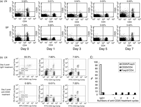
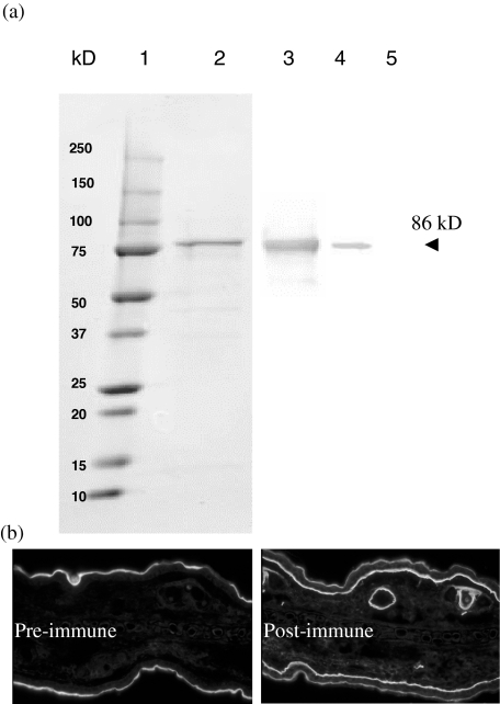
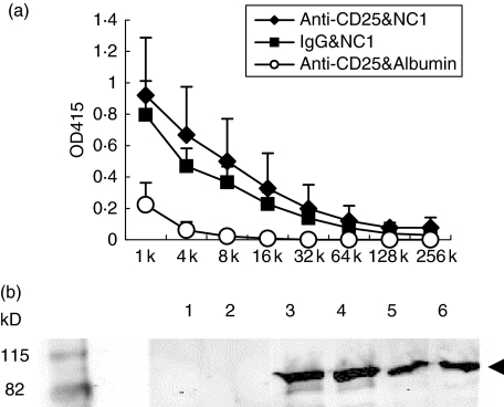
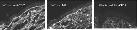
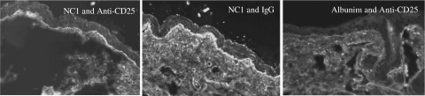
Epidermolysis bullosa acquisita is an autoimmune blistering disease characterized by circulating and skin basement membrane-bound IgG autoantibodies to type VII collagen, a major structural protein of the dermal-epidermal junction. Regulatory T cells (T(reg)) suppress self antigen-mediated autoimmune responses. To investigate the role of T(reg) in the the autoimmune response to type VII collagen in a mouse model, a monoclonal antibody against mouse CD25 was used to deplete T(reg). A recombinant mouse type VII collagen NC1 domain protein and mouse albumin were used as antigens. SKH1 mice were used as a testing host. Group 1 mice received NC1 immunization and were functionally depleted of T(reg); group 2 mice received NC1 immunization and rat isotype control; and group 3 mice received albumin immunization and were functionally depleted of T(reg). Results demonstrated that anti-NC1 IgG autoantibodies with high titres, as determined by enzyme-linked immunosorbent assay and Western blotting, developed in all mice immunized with NC1 (groups 1 and 2), but were undetected in group 3 mice. The predominant subclasses of anti-NC1 autoantibodies were IgG1, IgG2a and IgG2b; furthermore, these antibodies carried only the kappa light chain. IgG autoantibodies in the sera of NC1-immunized mice reacted with mouse skin basement membrane in vitro and deposited in skin basement membrane in vivo as detected by indirect and direct immunofluorescence microscopy, respectively. Our data suggest that the development of autoimmunity against type VII collagen in mice is independent of T(reg) function and the autoimmune response is mediated by both Th1 and Th2 cells. We speculate that the basement membrane deposition of IgG may eventually lead to blister development.


Figures

Fig. 1
Fig. 1
Effect of anti-CD25 monoclonal antibody. (a) SKH1 mice were intraperitoneally administered 500 µg purified rat anti-CD25. The mice which received the antibodies were killed at 24 h (day 1), day 3, day 5 or day 7 after the administration of the antibody. Spleen and lymph nodes were then harvested. Single-cell suspensions were stained with fluorescein isothiocyanate (FITC)-conjugated anti-mouse CD4 and phycoerythrin (PE)-labelled anti-mouse CD25 and analysed by fluorescence activated cell sorter (FACSCalibur). Results of injection of isotype rat Ig G are not shown. Day 0 indicates results of mice without anti-CD25 treatment. The numbers located above dot plots indicate the percentages of CD25+ cells in CD4+ cells. (b) To determine the expression of CD25 and forkhead boxp3 (Foxp3) in CD4+ cells after anti-CD25 antibody treatment, five groups of SKH1 mice were used. Group 1 received a single dose of 500 µg isotype IgG. Group 2 received a single dose of 500 µg anti-CD25. Group 3 received two doses of 500 µg anti-CD25, 5 days apart. Group 4 received three doses of 500 µg anti-CD25, 5 days apart. Group 5 received four doses of 500 µg anti-CD25, 5 days apart. At day 5 of the last injections, cells from the spleen and lymph node were stained by FITC-anti-CD4, PE-anti-Foxp3 and allophycocyanin (APC)-anti-CD25 and analysed by Cyan ADP flow cytometer. Data shown are results from lymph node cells in groups 1 and 2 after one anti-CD25 or IgG isotype injection. The numbers located above each dot plot in the left column indicate the percentages of CD25+ in Foxp3+ cells gated on CD4+ cells. The numbers located above each dot plot in the middle column indicate the percentages of CD25+ cells in CD4+ cells. The numbers located above each dot plot in the right column indicate the percentages of Foxp3+ cells in CD4+ cells. (c) Anti-CD25 inactivates Treg in a consistent manner: CD25+/Foxp3 (gated on CD4+), CD25+/CD4+ and Foxp3+/CD4+ cells in the lymph node were analysed after one, two, three and four cycles of anti-CD25 treatment, showing consistent inactivation of Treg, without physical depletion. Similar analyses for IgG1 isotype treatment obtained after one cycle treatment are shown on the far left for comparison. Similar results were obtained in the studies of the spleen cells (data not shown).
Fig. 2
Fig. 2
Generation of recombinant mouse type VII collagen NC1 domain and polyclonal antibodies against mouse NC1. (a). The cDNA of mouse NC1 domain encoding 580 amino acids at the N-terminal was cloned and expressed in Escherichia coli with a molecular mass of approximately 86 kD (lane 2, Coomassie blue-stained). The recombinant protein has a V5 epitope which is used for identifying the correct recombinant protein production by antibody to V5 (lane 3). The serum from the purified NC1 recombinant protein-immunized rat reacts specifically with NC1 (lane 4), while pre-immune serum (same dilution) of the same rat does not react with the protein (lane 5); 2·4 µg, 1·2 µg, 0·12 µg and 0·12 µg recombinant NC1 were loaded to lanes 2, 3, 4 and 5, respectively. (b). The serum from the purified NC1 recombinant protein-immunized rat also reacts distinctively with skin basement membrane zone of the dermal–epidermal junction and hair follicles (right panel), but the pre-immune serum of the same rat does not label it (left panel).
Fig. 3
Fig. 3
NC1-immunized SKH1 mice produce IgG antoantibodies specifically against recombinant NC1 protein. (a). Titratable enzyme-linked immunosorbent assay (ELISA) titres of autoantibodies. Three groups of female SKH1 mice were included. One group received rat anti-CD25 and mouse NC1 (n = 5, solid diamond), the second group received rat isotype and mouse NC1 (n = 5, solid square) and the third group received rat anti-CD25 and mouse albumin (n = 3, open circle). Mice sera were collected at the end of 5·5 months after the initial immunization and tested for the specific IgG against mouse NC1 by ELISA as described in Materials and methods. (b). Western blot analysis of NC1-specific autoantibodies. Two representative samples from each group are shown: mice treated with anti-CD25 and immunized with albumin (lanes 1 and 2); mice treated with anti-CD25 and immunized with NC1 (lanes 3 and 4); mice treated with rat IgG and immunized with NC1 (lanes 5 and 6). Each of the wells were loaded with 300 ng of recombinant mouse NC1 protein.
Fig. 4
Fig. 4
NC1-immunized SKH1 mice produce IgG autoantibodies which react specifically with skin basement membrane. Indirect immunofluorescence microscopy was used to examine the reactivity of autoreactive IgGs with the skin basement membrane zone component, as described in Materials and methods. Left and middle panels show IgG autoantibodies reacting with the skin basement membrane in mice immunized with NC1 in combinations of anti-CD25 or isotype control, respectively. The right panel shows the result of the serum from mouse treated with anti-CD25 and immunized with albumin. Images are representative for each group (original magnification ×40).
Fig. 5
Fig. 5
IgG class autoantibodies deposited to skin basement membrane in NC1-immunized mice. Direct immunofluorescence microscopy was performed to examine the deposit of IgG skin basement membrane zone, as described in Materials and methods. The left panel shows IgG deposits in mice treated with anti-CD25 and immunized with NC1, the middle panel shows IgG deposits in mice treated with rat IgG and immunized with NC1 and the right panel shows IgG deposits in mice treated with anti-CD25 and immunized with albumin. Images are representative for each group (original magnification ×40).

Similar articles

Cited by

References

    1. Woodley DT, Briggaman RA, O’Keefe EJ, Inman AO, Queen LL, Gammon WR. Identification of the skin basement-membrane autoantigen in epidermolysis bullosa acquisita. N Engl J Med. 1984;310:1007–13. - PubMed
    1. Yancey KB. The pathophysiology of autoimmune blistering diseases. J Clin Invest. 2005;115:825–8. - PMC - PubMed
    1. Chen M, Chan LS, Cai X, O'Toole EA, Sample JC, Woodley DT. Development of an ELISA for rapid detection of anti-type VII collagen autoantibodies in epidermolysis bullosa acquisita. J Invest Dermatol. 1997;108:68–72. - PubMed
    1. Chen M, Keene DR, Costa FK, Tahk SH, Woodley DT. The carboxyl terminus of type VII collagen mediates antiparallel dimer formation and constitutes a new antigenic epitope for epidermolysis bullosa acquisita autoantibodies. J Biol Chem. 2001;276:21649–55. - PubMed
    1. Lapiere JC, Woodley DT, Parente MG, et al. Epitope mapping of type VII collagen. Identification of discrete peptide sequences recognized by sera from patients with acquired epidermolysis bullosa. J Clin Invest. 1993;92:1831–9. - PMC - PubMed

Publication types

MeSH terms