

Regulation of bladder muscarinic receptor subtypes by experimental pathologies
- PMID: 16879497
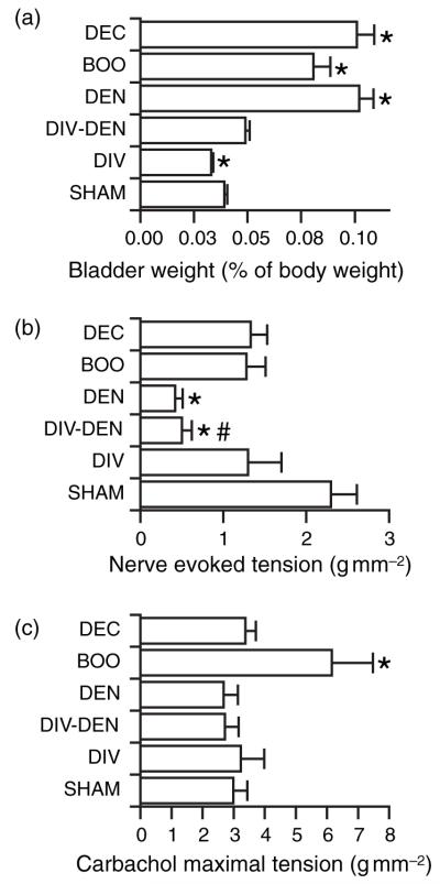
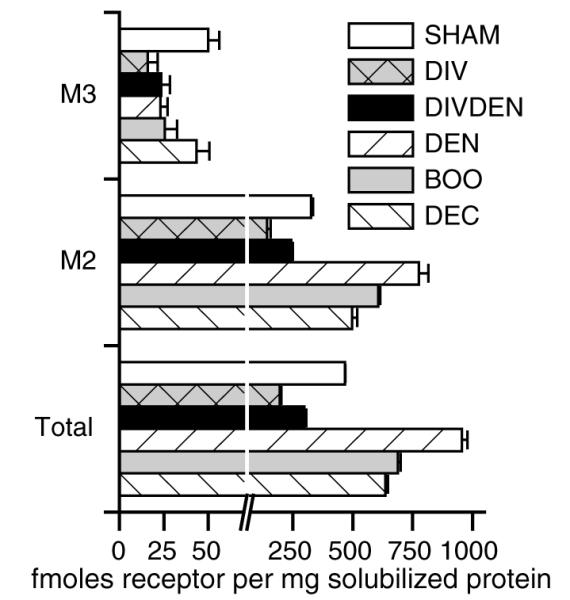
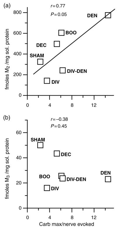
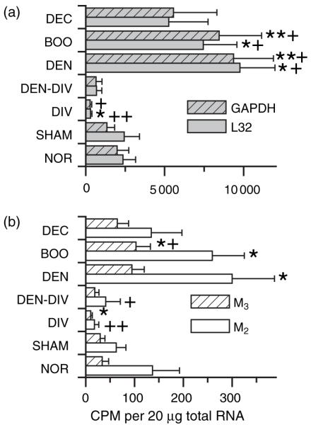
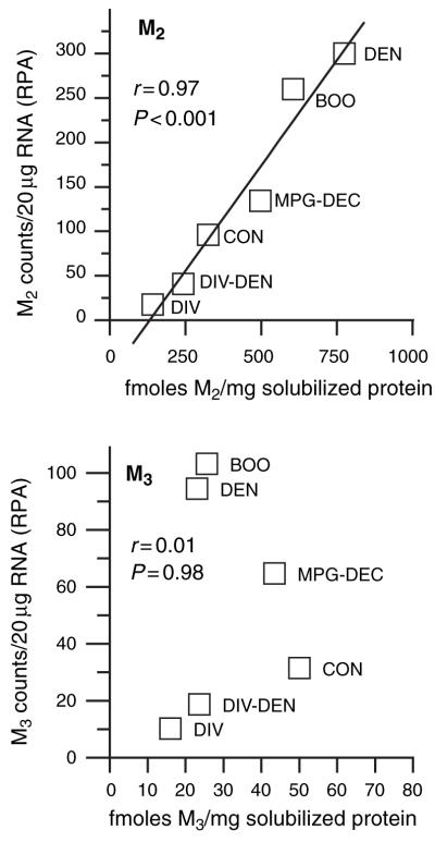
- PMCID: PMC3275807
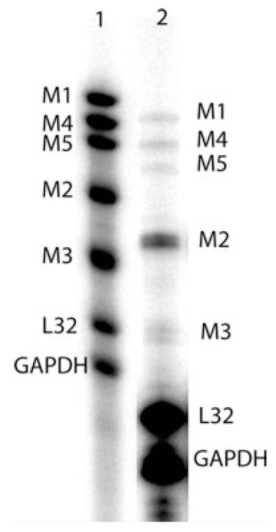
- DOI: 10.1111/j.1474-8673.2006.00377.x
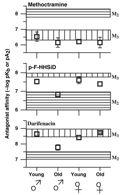
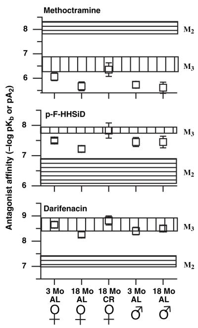
Regulation of bladder muscarinic receptor subtypes by experimental pathologies
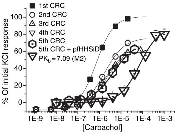
Abstract
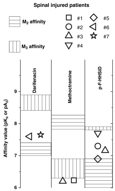
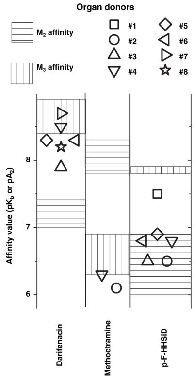
1 The M3 muscarinic receptor subtype is widely accepted as the receptor on smooth muscle cells that mediates cholinergic contraction of the normal urinary bladder and other smooth muscle tissues, however, we have found that the M2 receptor participates in contraction under certain abnormal conditions. The aim of this study was to determine the effects of various experimental pathologies on the muscarinic receptor subtype mediating urinary bladder contraction. 2 Experimental pathologies resulting in bladder hypertrophy (denervation and outlet obstruction) result in an up-regulation of bladder M2 receptors and a change in the receptor subtype mediating contraction from M3 towards M2. Preventing the denervation-induced bladder hypertrophy by urinary diversion prevents this shift in contractile phenotype indicating that hypertrophy is responsible as opposed to denervation per se. 3 The hypertrophy-induced increase in M2 receptor density and contractile response is accompanied by an increase in the tissue concentrations of mRNA coding for the M2 receptor subtype, however, M3 receptor protein density does not correlate with changes in M3 receptor tissue mRNA concentrations across different experimental pathologies. 4 This shift in contractile phenotype from M3 towards M2 subtype is also observed in aged male Sprague-Dawley rats but not females or either sex of the Fisher344 strain of rats. 5 Four repeated, sequential agonist concentration response curves also cause this shift in contractile phenotype in normal rat bladder strips in vitro, as evidenced by a decrease in the affinity of the M3 selective antagonist p-fluoro-hexahydro-sila-diphenidol (p-F-HHSiD). 6 A similar decrease in the contractile affinity of M3 selective antagonists (darifenacin and p-F-HHSiD) is also observed in bladder specimens from patients with neurogenic bladder as well as certain organ transplant donors. 7 It is concluded that although the M3 receptor subtype predominantly mediates contraction under normal circumstances, the M2 receptor subtype can take over a contractile role when the M3 subtype becomes inactivated by, for example, repeated agonist exposures or bladder hypertrophy. This finding has substantial implications for the clinical treatment of abnormal bladder contractions.
Figures
Similar articles
-
Hypertrophy changes the muscarinic receptor subtype mediating bladder contraction from M3 toward M2.Am J Physiol Regul Integr Comp Physiol. 2003 Sep;285(3):R701-8. doi: 10.1152/ajpregu.00009.2003. Epub 2003 May 22. Am J Physiol Regul Integr Comp Physiol. 2003. PMID: 12763741 Free PMC article.
-
M2 and M3 muscarinic receptor activation of urinary bladder contractile signal transduction. II. Denervated rat bladder.J Pharmacol Exp Ther. 2006 Feb;316(2):875-80. doi: 10.1124/jpet.105.094961. Epub 2005 Oct 21. J Pharmacol Exp Ther. 2006. PMID: 16243962
-
M2 receptors in genito-urinary smooth muscle pathology.Life Sci. 1999;64(6-7):429-36. doi: 10.1016/s0024-3205(98)00582-7. Life Sci. 1999. PMID: 10069506 Free PMC article.
-
Muscarinic receptor subtypes modulating smooth muscle contractility in the urinary bladder.Life Sci. 1999;64(6-7):419-28. doi: 10.1016/s0024-3205(98)00581-5. Life Sci. 1999. PMID: 10069505 Review.
-
Contractile role of M2 and M3 muscarinic receptors in gastrointestinal smooth muscle.Life Sci. 1999;64(6-7):387-94. doi: 10.1016/s0024-3205(98)00584-0. Life Sci. 1999. PMID: 10069501 Review.
Cited by
-
Perspectives on overactive bladder in the elderly population.World J Urol. 2009 Dec;27(6):729-37. doi: 10.1007/s00345-009-0491-0. Epub 2009 Nov 11. World J Urol. 2009. PMID: 19904542 Review.
-
Alterations in nerve-evoked bladder contractions in a coronavirus-induced mouse model of multiple sclerosis.PLoS One. 2014 Oct 13;9(10):e109314. doi: 10.1371/journal.pone.0109314. eCollection 2014. PLoS One. 2014. PMID: 25310403 Free PMC article.
-
The role of the mucosa in modulation of evoked responses in the spinal cord injured rat bladder.Neurourol Urodyn. 2018 Jun;37(5):1583-1593. doi: 10.1002/nau.23512. Epub 2018 Feb 10. Neurourol Urodyn. 2018. PMID: 29427331 Free PMC article.
-
Impairment of ATP hydrolysis decreases adenosine A1 receptor tonus favoring cholinergic nerve hyperactivity in the obstructed human urinary bladder.Purinergic Signal. 2015 Dec;11(4):595-606. doi: 10.1007/s11302-015-9478-z. Epub 2015 Oct 31. Purinergic Signal. 2015. PMID: 26521170 Free PMC article.
-
Activation of muscarinic M3 receptors inhibits large-conductance voltage- and Ca2+-activated K+ channels in rat urinary bladder smooth muscle cells.Am J Physiol Cell Physiol. 2013 Jul 15;305(2):C207-14. doi: 10.1152/ajpcell.00113.2013. Epub 2013 May 22. Am J Physiol Cell Physiol. 2013. PMID: 23703523 Free PMC article.
References
-
- ABRAMS P. Evidence for the efficacy and safety of tolterodine in the treatment of overactive bladder. Exp. Opinion Pharmacothera. 2001;2:1685–1701. - PubMed
-
- ANDERSSON KE, HOLMQUIST F, FOVAEUS M, HEDLUND H, SUNDLER R. Muscarinic receptor stimulation of phosphoinositide hydrolysis in the human isolated urinary bladder. J. Urol. 1991;146:1156–1159. - PubMed
-
- BAS A, FORSBERG G, HAMMARSTROM S, HAMMAR-STROM ML. Utility of the housekeeping genes 18S rRNA, beta-actin and glyceraldehyde-3-phosphate-dehydrogenase for normalization in real-time quantitative reverse transcriptase-polymerase chain reaction analysis of gene expression in human T lymphocytes. Scandi. J. Immunol. 2004;59:566–573. - PubMed
-
- BATRA S, BIORKLUND A, HEDLUND H, ANDERSSON KE, BJORKLUND A. Identification and characterization of muscarinic cholinergic receptors in the human urinary bladder and parotid gland. J. Auton. Nerv. Syst. 1988;22:174. 1987. [erratum appears. Note: Bjorklund A (corrected to Biorklund A)]. J. Auton. Nerv. Syst., 20, 129-135. - PubMed
-
- BAYLISS M, WU C, NEWGREEN D, MUNDY AR, FRY CH. A quantitative study of atropine-resistant contractile responses in human detrusor smooth muscle, from stable, unstable and obstructed bladders. J. Urol. 1999;162:1833–1839. - PubMed
MeSH terms
Substances
Grants and funding
LinkOut - more resources
Full Text Sources
