

Mechanisms of critical illness--classifying microcirculatory flow abnormalities in distributive shock
- PMID: 16879732
- PMCID: PMC1750971
- DOI: 10.1186/cc4969
Mechanisms of critical illness--classifying microcirculatory flow abnormalities in distributive shock
Abstract
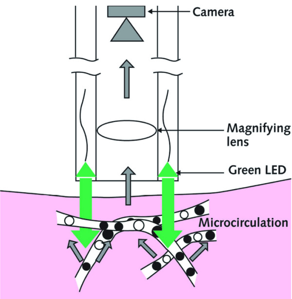
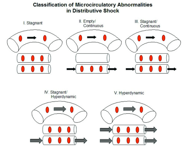
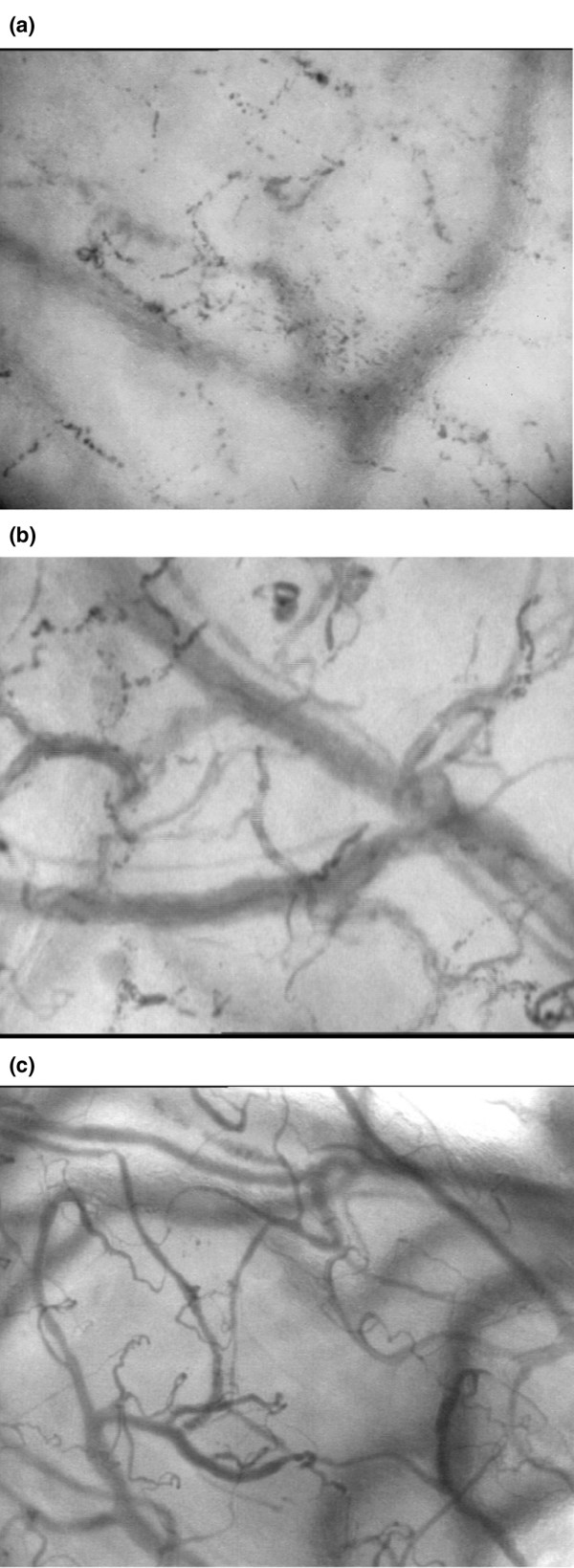
Over 30 years ago Weil and Shubin proposed a re-classification of shock states and identified hypovolemic, cardiogenic, obstructive and distributive shock. The first three categories have in common that they are associated with a fall in cardiac output. Distributive shock, such as occurs during sepsis and septic shock, however, is associated with an abnormal distribution of microvascular blood flow and metabolic distress in the presence of normal or even supranormal levels of cardiac output. This Bench-to-bedside review looks at the recent insights that have been gained into the nature of distributive shock. Its pathophysiology can best be described as a microcirculatory and mitochondrial distress syndrome, where time and therapy form an integral part of the definition. The clinical introduction of new microcirculatory imaging techniques, such as orthogonal polarization spectral and side-stream dark-field imaging, have allowed direct observation of the microcirculation at the bedside. Images of the sublingual microcirculation during septic shock and resuscitation have revealed that the distributive defect of blood flow occurs at the capillary level. In this paper, we classify the different types of heterogeneous flow patterns of microcirculatory abnormalities found during different types of distributive shock. Analysis of these patterns gave a five class classification system to define the types of microcirculatory abnormalities found in different types of distributive shock and indicated that distributive shock occurs in many other clinical conditions than just sepsis and septic shock. It is likely that different mechanisms defined by pathology and treatment underlie these abnormalities observed in the different classes. Functionally, however, they all cause a distributive defect resulting in microcirculatory shunting and regional dysoxia. It is hoped that this classification system will help in the identification of mechanisms underlying these abnormalities and indicate optimal therapies for resuscitating septic and other types of distributive shock.
Figures
Similar articles
-
Similar Microcirculatory Alterations in Patients with Normodynamic and Hyperdynamic Septic Shock.Ann Am Thorac Soc. 2016 Feb;13(2):240-7. doi: 10.1513/AnnalsATS.201509-606OC. Ann Am Thorac Soc. 2016. PMID: 26624559
-
Disparity between skin perfusion and sublingual microcirculatory alterations in severe sepsis and septic shock: a prospective observational study.Intensive Care Med. 2008 Jul;34(7):1294-8. doi: 10.1007/s00134-008-1007-x. Epub 2008 Mar 4. Intensive Care Med. 2008. PMID: 18317733 Free PMC article. Clinical Trial.
-
The heterogeneity of the microcirculation in critical illness.Clin Chest Med. 2008 Dec;29(4):643-54, viii. doi: 10.1016/j.ccm.2008.06.008. Clin Chest Med. 2008. PMID: 18954699 Review.
-
Early microcirculatory perfusion derangements in patients with severe sepsis and septic shock: relationship to hemodynamics, oxygen transport, and survival.Ann Emerg Med. 2007 Jan;49(1):88-98, 98.e1-2. doi: 10.1016/j.annemergmed.2006.08.021. Epub 2006 Nov 7. Ann Emerg Med. 2007. PMID: 17095120
-
What is microcirculatory shock?Curr Opin Crit Care. 2015 Jun;21(3):245-52. doi: 10.1097/MCC.0000000000000196. Curr Opin Crit Care. 2015. PMID: 25827583 Review.
Cited by
-
SOFA Score, Hemodynamics and Body Temperature Allow Early Discrimination between Porcine Peritonitis-Induced Sepsis and Peritonitis-Induced Septic Shock.J Pers Med. 2021 Feb 28;11(3):164. doi: 10.3390/jpm11030164. J Pers Med. 2021. PMID: 33670874 Free PMC article.
-
Comparison of two different generations of "NIRS" devices and transducers in healthy volunteers and ICU patients.J Clin Monit Comput. 2013 Feb;27(1):71-9. doi: 10.1007/s10877-012-9400-y. Epub 2012 Oct 7. J Clin Monit Comput. 2013. PMID: 23054384
-
Is the Sympathetic System Detrimental in the Setting of Septic Shock, with Antihypertensive Agents as a Counterintuitive Approach? A Clinical Proposition.J Clin Med. 2021 Oct 1;10(19):4569. doi: 10.3390/jcm10194569. J Clin Med. 2021. PMID: 34640590 Free PMC article. Review.
-
A guide to human in vivo microcirculatory flow image analysis.Crit Care. 2016 Feb 10;20:35. doi: 10.1186/s13054-016-1213-9. Crit Care. 2016. PMID: 26861691 Free PMC article.
-
The dynamic regulation of microcirculatory conduit function: features relevant to transfusion medicine.Transfus Apher Sci. 2010 Aug;43(1):61-8. doi: 10.1016/j.transci.2010.05.010. Epub 2010 Jun 26. Transfus Apher Sci. 2010. PMID: 20580315 Free PMC article. Review.
References
-
- Weil MH, Shubin H. Proposed reclassification of shock states with special reference to distributive defects. Adv Exp Med Biol. 1971;23:13–23. - PubMed
-
- Siegemund M, van Bommel J, Schwarte LA, Studer W, Girard T, Marsch S, Radermacher P, Ince C. Inducible nitric oxide synthase inhibition improves intestinal microcirculatory oxygenation and CO2 balance during endotoxemia in pigs. Intensive Care Med. 2005;31:985–992. doi: 10.1007/s00134-005-2664-7. - DOI - PubMed
Publication types
MeSH terms
LinkOut - more resources
Full Text Sources
Other Literature Sources
Medical
