

Characterisation of the immune response to type I collagen in scleroderma
- PMID: 16879746
- PMCID: PMC1779396
- DOI: 10.1186/ar2025
Characterisation of the immune response to type I collagen in scleroderma
Abstract
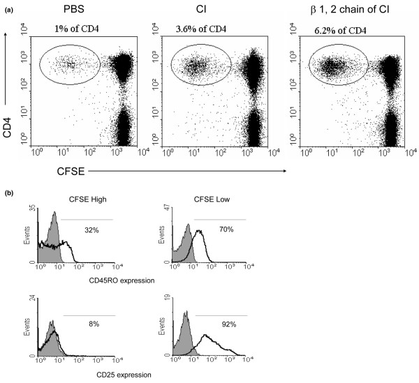
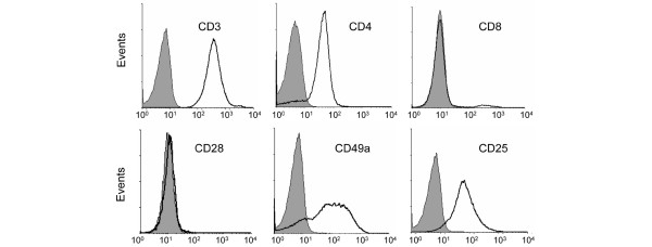
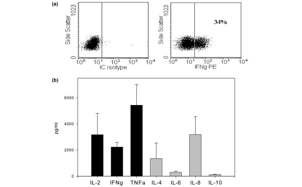
This study was conducted to examine the frequency, phenotype, and functional profile of T lymphocytes that proliferate in response to type I collagen (CI) in patients with scleroderma (SSc). Peripheral blood mononuclear cells (PBMCs) from SSc patients, healthy controls, and rheumatoid arthritis disease controls were labeled with carboxy-fluorescein diacetate, succinimidyl ester (CFSE), cultured with or without antigen (bovine CI) for 14 days, and analysed by flow cytometry. Surface markers of proliferating cells were identified by multi-color flow cytometry. T-cell lines were derived after sorting for proliferating T cells (CFSElow). Cytokine expression in CI-responsive T cells was detected by intracellular staining/flow cytometry and by multiplex cytokine bead assay (Bio-Plex). A T-cell proliferative response to CI was detected in 8 of 25 (32%) SSc patients, but was infrequent in healthy or disease controls (3.6%; p = 0.009). The proliferating T cells expressed a CD4+, activated (CD25+), memory (CD45RO+) phenotype. Proliferation to CI did not correlate with disease duration or extent of skin involvement. T-cell lines were generated using in vitro CI stimulation to study the functional profile of these cells. Following activation of CI-reactive T cells, we detected intracellular interferon (IFN)-gamma but not interleukin (IL)-4 by flow cytometry. Supernatants from the T-cell lines generated in vitro contained IL-2, IFN-gamma, GM-CSF (granulocyte macrophage-colony-stimulating factor), and tumour necrosis factor-alpha, but little or no IL-4 and IL-10, suggesting that CI-responsive T cells express a predominantly Th1 cytokine pattern. In conclusion, circulating memory CD4 T cells that proliferate to CI are present in a subset of patients with SSc, but are infrequent in healthy or disease controls.
Figures
Similar articles
-
Analysis of Th1 and Th2 cytokines expressing CD4+ and CD8+ T cells in rheumatoid arthritis by flow cytometry.J Rheumatol. 2000 May;27(5):1128-35. J Rheumatol. 2000. PMID: 10813277
-
Peripheral blood CD4 but not CD8 t-lymphocytes in patients with exacerbation of asthma transcribe and translate messenger RNA encoding cytokines which prolong eosinophil survival in the context of a Th2-type pattern: effect of glucocorticoid therapy.Am J Respir Cell Mol Biol. 1995 May;12(5):567-78. doi: 10.1165/ajrcmb.12.5.7742019. Am J Respir Cell Mol Biol. 1995. PMID: 7742019
-
Systemic sclerosis Th2 cells inhibit collagen production by dermal fibroblasts via membrane-associated tumor necrosis factor alpha.Arthritis Rheum. 2003 Sep;48(9):2593-604. doi: 10.1002/art.11129. Arthritis Rheum. 2003. PMID: 13130479
-
T and NK Cell Phenotypic Abnormalities in Systemic Sclerosis: a Cohort Study and a Comprehensive Literature Review.Clin Rev Allergy Immunol. 2015 Dec;49(3):347-69. doi: 10.1007/s12016-015-8505-8. Clin Rev Allergy Immunol. 2015. PMID: 26445774 Review.
-
Altered B lymphocyte homeostasis and functions in systemic sclerosis.Autoimmun Rev. 2018 Mar;17(3):244-255. doi: 10.1016/j.autrev.2017.10.015. Epub 2018 Jan 16. Autoimmun Rev. 2018. PMID: 29343447 Review.
Cited by
-
Pharmaceutical Applications of Biomass Polymers: Review of Current Research and Perspectives.Polymers (Basel). 2024 Apr 23;16(9):1182. doi: 10.3390/polym16091182. Polymers (Basel). 2024. PMID: 38732651 Free PMC article. Review.
-
Stimulation with type I collagen induces changes in gene expression in peripheral blood mononuclear cells from patients with diffuse cutaneous systemic sclerosis (scleroderma).Clin Exp Immunol. 2010 Sep;161(3):426-35. doi: 10.1111/j.1365-2249.2010.04189.x. Clin Exp Immunol. 2010. PMID: 20529088 Free PMC article.
-
Supernatants from culture of type I collagen-stimulated PBMC from patients with cutaneous systemic sclerosis versus localized scleroderma demonstrate suppression of MMP-1 by fibroblasts.Clin Rheumatol. 2012 Jun;31(6):973-81. doi: 10.1007/s10067-012-1962-z. Epub 2012 Feb 25. Clin Rheumatol. 2012. PMID: 22367096 Free PMC article.
-
Pathogenesis of Systemic Sclerosis.Front Immunol. 2015 Jun 8;6:272. doi: 10.3389/fimmu.2015.00272. eCollection 2015. Front Immunol. 2015. PMID: 26106387 Free PMC article. Review.
-
Vascular involvement in systemic sclerosis (scleroderma).J Inflamm Res. 2011;4:105-25. doi: 10.2147/JIR.S18145. Epub 2011 Jul 26. J Inflamm Res. 2011. PMID: 22096374 Free PMC article.
References
-
- Seyer JM, Kang AH, Rodnan G. Investigation of type I and type III collagens of the lung in progressive systemic sclerosis. Arthritis Rheum. 1981;24:625–631. - PubMed
-
- Steen VD, Engel EE, Charley MR, Medsger TA., Jr Soluble serum interleukin 2 receptors in patients with systemic sclerosis. J Rheumatol. 1996;23:646–649. - PubMed
Publication types
MeSH terms
Substances
Grants and funding
LinkOut - more resources
Full Text Sources
Other Literature Sources
Medical
Research Materials
