

Integrative roles of transforming growth factor-alpha in the cytoprotection mechanisms of gastric mucosal injury
- PMID: 16879752
- PMCID: PMC1552080
- DOI: 10.1186/1471-230X-6-22
Integrative roles of transforming growth factor-alpha in the cytoprotection mechanisms of gastric mucosal injury
Abstract
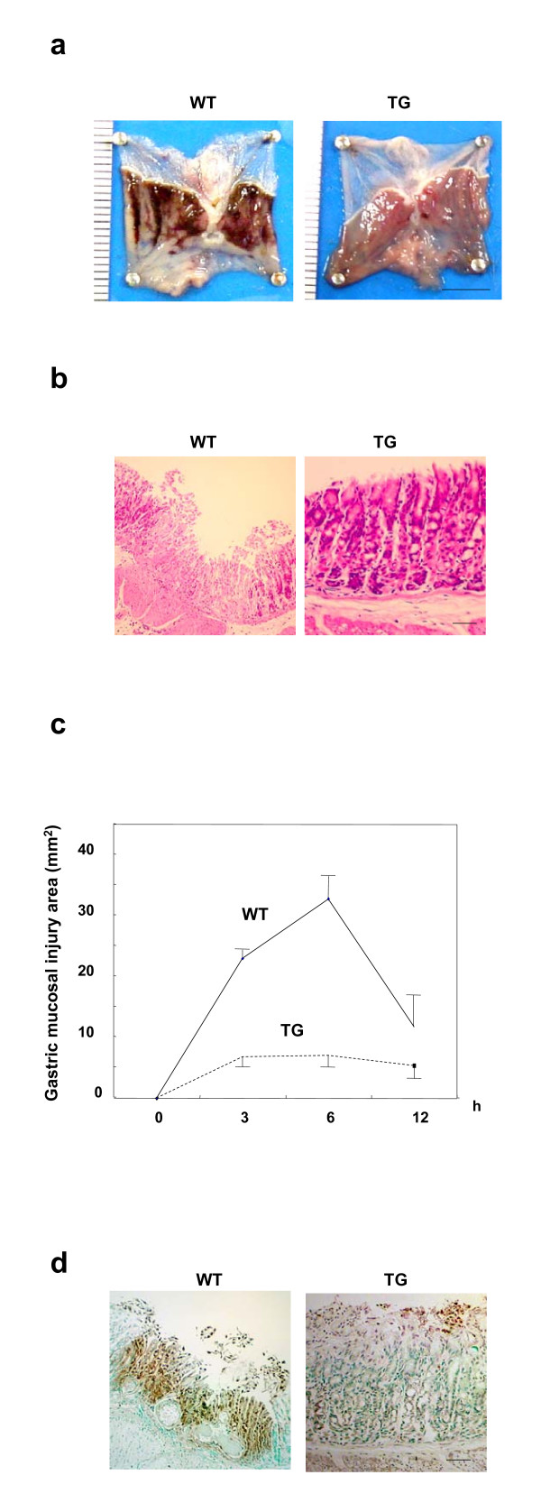
Background: Transforming growth factor alpha (TGFalpha) protects against gastric mucosal injury and facilitates wound healing. However, its overexpression is known to induce hypertrophic gastropathy resembling Menetrier's disease in transgenic (TG) mice on an FVB background, as one of the authors reported previously. We studied another TGFalpha-expressing mouse line on a CD1 background, whose gastric mucosa appears normal. Since this TG mouse had a strong resistance to ethanol-induced gastric injury, we considered the long-term effect of TGFalpha on several gastric protection mechanisms.
Methods: TGFalpha-expressing transgenic (TG) mouse lines bearing human TGFalpha cDNA under the control of the mouse metallothionein gene I promoter were generated on a CD1 mouse background, and analyzed their ethanol injury-resistant phenotypes produced by TGFalpha.
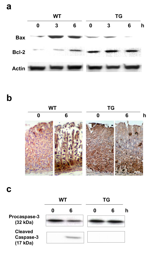
Results: In the TG mucosa, blood flow was well maintained after ethanol injury. Further, neural and inducible types of NO synthases were consistently and widely expressed in the TG mucosa, compared with the limited distribution of neural type NO synthase in the luminal pit region of the wild-type (WT) mucosa. COX-2 and its upstream transcription factor NfkB were constitutively elevated in the TG mucosa even before ethanol administration, whereas they were induced in the same region of the WT mucosa only after ethanol injury. Two anti-apoptotic proteins, HSP70 and Bcl-2, were upregulated in the TG mucosa even before ethanol administration, while they were not expressed in the WT mucosa before the injury. Furthermore, pro-caspase 3 activation was inhibited in the TG mucosa, while it was converted to the active form in the WT mucosa following ethanol administration.
Conclusion: We conclude that TGFalpha maintains the gastric mucosal defense against gastric injury by integrating other cytoprotective mechanisms.
Figures


Similar articles
-
Ghrelin-a new gastroprotective factor in gastric mucosa.J Physiol Pharmacol. 2004 Jun;55(2):325-36. J Physiol Pharmacol. 2004. PMID: 15213356
-
Protective role of adiponectin against ethanol-induced gastric injury in mice.Am J Physiol Gastrointest Liver Physiol. 2012 Apr 15;302(8):G773-80. doi: 10.1152/ajpgi.00324.2011. Epub 2012 Feb 9. Am J Physiol Gastrointest Liver Physiol. 2012. PMID: 22323129
-
Cyclooxygenase 2, pS2, inducible nitric oxide synthase and transforming growth factor alpha in gastric adaptation to stress.World J Gastroenterol. 2004 Dec 1;10(23):3537-41. doi: 10.3748/wjg.v10.i23.3537. World J Gastroenterol. 2004. PMID: 15526382 Free PMC article.
-
Neural aspects of prostaglandin involvement in gastric mucosal defense.J Physiol Pharmacol. 2001 Dec;52(4 Pt 1):555-68. J Physiol Pharmacol. 2001. PMID: 11787758 Review.
-
Roles for transforming growth factor-alpha in gastric physiology and pathophysiology.Yale J Biol Med. 1992 Nov-Dec;65(6):693-704; discussion 621-3. Yale J Biol Med. 1992. PMID: 1341072 Free PMC article. Review.
Cited by
-
Ménétrier disease and gastrointestinal stromal tumors: hyperproliferative disorders of the stomach.J Clin Invest. 2007 Jan;117(1):70-80. doi: 10.1172/JCI30491. J Clin Invest. 2007. PMID: 17200708 Free PMC article. Review.
-
Gut homeostasis, injury, and healing: New therapeutic targets.World J Gastroenterol. 2022 May 7;28(17):1725-1750. doi: 10.3748/wjg.v28.i17.1725. World J Gastroenterol. 2022. PMID: 35633906 Free PMC article. Review.
-
Hepatocyte growth factor overexpression ameliorates liver inflammation and fibrosis in a mouse model of nonalcoholic steatohepatitis.Hepatol Int. 2012 Jun;6(3):620-30. doi: 10.1007/s12072-011-9301-z. Epub 2011 Aug 5. Hepatol Int. 2012. PMID: 21818687
-
Annexin A1, formyl peptide receptor, and NOX1 orchestrate epithelial repair.J Clin Invest. 2013 Jan;123(1):443-54. doi: 10.1172/JCI65831. Epub 2012 Dec 17. J Clin Invest. 2013. PMID: 23241962 Free PMC article.
References
-
- Kamimura H, Konda Y, Yokota H, Takenoshita S, Nagamachi Y, Kuwano H, Takeuchi T. Kex2 family endoprotease furin is expressed specifically in pit-region parietal cells of the rat gastric mucosa. Am J Physiol. 1999;277:G183–G190. - PubMed
-
- Nakajima T, Konda Y, Izumi Y, Kanai M, Hayashi N, Chiba T, Takeuchi T. Gastrin stimulates the growth of gastric pit cell precursors by inducing its own receptors. Am J Physiol Gastrointest Liver Physiol. 2002;282:G359–G366. - PubMed
Publication types
MeSH terms
Substances
LinkOut - more resources
Full Text Sources
Medical
Research Materials
Miscellaneous
