. 2006 Aug 7;203(8):2009-19.
doi: 10.1084/jem.20052222. Epub 2006 Jul 31.

T-bet negatively regulates autoimmune myocarditis by suppressing local production of interleukin 17

Affiliations

T-bet negatively regulates autoimmune myocarditis by suppressing local production of interleukin 17

Manu Rangachari et al. J Exp Med. .

Abstract

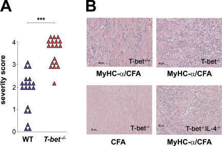
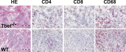
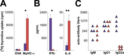
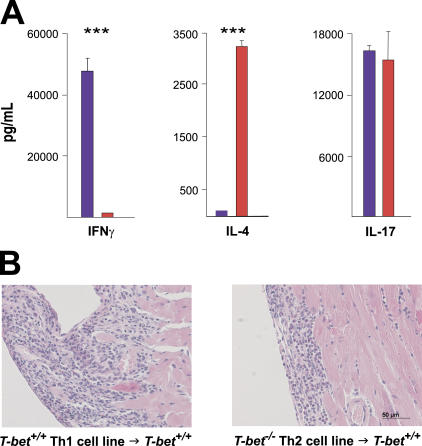
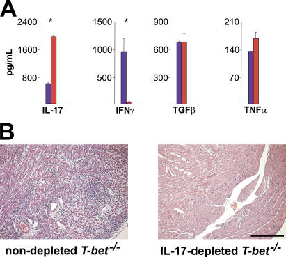
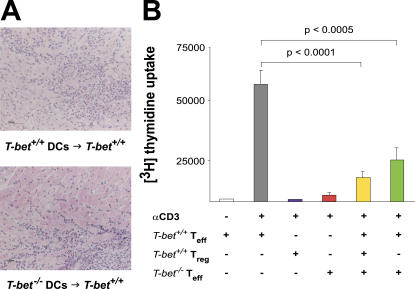
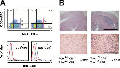
Experimental autoimmune myocarditis (EAM) appears after infectious heart disease, the most common cause of dilated cardiomyopathy in humans. Here we report that mice lacking T-bet, a T-box transcription factor required for T helper (Th)1 cell differentiation and interferon (IFN)-gamma production, develop severe autoimmune heart disease compared to T-bet+/+ control mice. Experiments in T-bet-/- IL-4-/- and T-bet-/- IL-4Ralpha-/- mice, as well as transfer of heart-specific Th1 and Th2 cell lines, showed that autoimmune heart disease develops independently of Th1 or Th2 polarization. Analysis of T-bet-/- IL-12Rbeta1-/- and T-bet-/- IL-12p35-/- mice then identified interleukin (IL)-23 as critical for EAM pathogenesis. In addition, T-bet-/- mice showed a marked increase in production of the IL-23-dependent cytokine IL-17 by heart-infiltrating lymphocytes, and in vivo IL-17 depletion markedly reduced EAM severity in T-bet-/- mice. Heart-infiltrating T-bet-/- CD8+ but not CD8- T cells secrete IFN-gamma, which inhibits IL-17 production and protects against severe EAM. In contrast, T-bet-/- CD8+ lymphocytes completely lost their capacity to release IFN-gamma within the heart. Collectively, these data show that severe IL-17-mediated EAM can develop in the absence of T-bet, and that T-bet can regulate autoimmunity via the control of nonspecific CD8+ T cell bystander functions in the inflamed target organ.


Figures

Figure 1.
Figure 1.
T-bet−/− mice develop EAM of increased severity. T-bet +/+ and T-bet −/− mice were immunized with MyHC-α peptide in CFA and killed 21 d later. (A) Severity scores of individual diseased T-bet +/+ versus T-bet −/− mice: ***, P < 0.000001. (B) Hematoxylin and eosin–stained sections from hearts of immunized mice. MyHC-α–immunized T-bet +/+ (top left), MyHC-α–immunized T-bet −/− (top right), T-bet −/− immunized with CFA alone (bottom left), and MyHC-α–immunized T-bet −/− IL-4 −/−.
Figure 2.
Figure 2.
Characterization of heart-infiltrating cells in T-bet−/− mice. Frozen heart sections from MyHC-α–immunized T-bet +/+ and T-bet −/− mice were stained to determine proportions of infiltrating macrophages (CD68, right) and lymphocytes (CD4, second from left; CD8, second from right).
Figure 3.
Figure 3.
CD4 T cell responses and cytokine production patterns of immunized T-bet−/− versus T-bet+/+ mice. CD4+ T cells from T-bet +/+ (blue bars) and T-bet −/− mice (red bars) were restimulated with either MyHC-α or control OVA peptide. (A) Proliferative responses after 48 h, and (B) IFN-γ and IL-4 production in supernatants after 40 h of restimulation, representative of three independent experiments. (C) Serum MyHC-α autoantibody responses from immunized T-bet +/+ (blue) and T-bet −/− (red) mice at day 21. *, P < 0.05; **, P < 0.01.
Figure 4.
Figure 4.
Th1 as well as Th2 CD4+ T cell lines trigger autoimmune myocarditis. (A) MyHC-α–specific lines were generated by subjecting CD4+ splenocytes from immunized T-bet +/+ (blue) or T-bet −/− (red) mice to multiple rounds of antigen restimulation followed by rest in minimal IL-2. Supernatant was collected for ELISA 48 h after the third antigen restimulation. ***, P < 0.0001. One representative experiment is shown. (B) Both MyHC-α–specific Th1 cell lines (left) and Th2 cell lines (right) are pathogenic in wild-type recipient mice. Hematoxylin and eosin–stained sections: bar: 50 μm.
Figure 5.
Figure 5.
Heart-infiltrating T-bet−/− CD3+ T cells display an altered cytokine profile, and IL-17 depletion improves EAM severity in T-bet−/− mice. (A) Cytokine production profile of T-bet −/− heart-infiltrating T cells. Infiltrating CD3+ cells were isolated from the hearts of immunized T-bet +/+ (blue) or T-bet −/− (red) mice and restimulated with anti-CD3/anti-CD28 for 24 h. Production of indicated cytokines was assessed by supernatant ELISA. *, P < 0.005. A representative of two individual experiments is shown. (B) T-bet −/− mice were immunized with 50 μg MyHC-α and injected with either 100 μg anti–mouse IL-17 or with 100 μl PBS on days 9, 12, and 15. Mice were killed on day 17. Hematoxylin and eosin–stained sections: bar, 200 μm.
Figure 6.
Figure 6.
T-bet−/− DCs and CD4+CD25+ T reg cells function normally. (A) T-bet +/+ mice were immunized with activated, MyHC-α–pulsed T-bet +/+ or T-bet −/− BMDCs. (B) T-bet +/+ CD4+CD25 T cells (Teff) were stimulated with anti-CD3 with or without T-bet +/+ or T-bet −/− CD4+CD25+ cells (Treg). Proliferation was measured at 72 h by [3H]thymidine incorporation. Data are representative of three independent experiments.
Figure 7.
Figure 7.
Heart-infiltrating T-bet−/− CD8+ T cells display impaired IFN-γ production, and loss of T-bet in CD8+ T cells exacerbates myocarditis. (A) Heart-infiltrating mononuclear cells from immunized mice were restimulated with anti-CD3/anti-CD28, stained for CD3, CD8, and intracellular IFN-γ, and analyzed by flow cytometry. Blue boxes and histograms, T-bet +/+; red boxes and histograms, T-bet −/−. A representative of two individual experiments is shown. (B) SCID mice were reconstituted with either T-bet +/+ CD4+ plus T-bet +/+ CD8+ or T-bet +/+ CD4+ plus T-bet −/− CD8+ and immunized. Mice were killed 21 d later. Hematoxylin and eosin–stained sections of hearts from mice receiving T-bet −/− CD8+ cells (right) versus mice receiving T-bet +/+ CD8+ cells (left) are shown. Bars, (top) 1 mm; (bottom) 100 μm.

Similar articles

Cited by

References

    1. Eriksson, U., and J.M. Penninger. 2005. Autoimmune heart failure: new understandings of pathogenesis. Int. J. Biochem. Cell Biol. 37:27–32. - PubMed
    1. Koelsch, S.P.S., G. Hufnagel, and B. Maisch. 2004. The European study of epidemiology and treatment of cardiac inflammatory diseases (ESETCID)—epidemiological results after 6 years. In Annual Meeting of the AHA, New Orleans.
    1. Caforio, A.L., E. Bonifacio, J.T. Stewart, D. Neglia, O. Parodi, G.F. Bottazzo, and W.J. McKenna. 1990. Novel organ-specific circulating cardiac autoantibodies in dilated cardiomyopathy. J. Am. Coll. Cardiol. 15:1527–1534. - PubMed
    1. Frustaci, A., C. Chimenti, F. Calabrese, M. Pieroni, G. Thiene, and A. Maseri. 2003. Immunosuppressive therapy for active lymphocytic myocarditis: virological and immunologic profile of responders versus nonresponders. Circulation. 107:857–863. - PubMed
    1. Omerovic, E., E. Bollano, B. Andersson, V. Kujacic, W. Schulze, A. Hjalmarson, F. Waagstein, and M. Fu. 2000. Induction of cardiomyopathy in severe combined immunodeficiency mice by transfer of lymphocytes from patients with idiopathic dilated cardiomyopathy. Autoimmunity. 32:271–280. - PubMed

Publication types

MeSH terms