. 2006 Aug 7;203(8):1883-9.
doi: 10.1084/jem.20060336. Epub 2006 Jul 31.

Antiinflammatory effects of dexamethasone are partly dependent on induction of dual specificity phosphatase 1

Affiliations

Antiinflammatory effects of dexamethasone are partly dependent on induction of dual specificity phosphatase 1

Sonya M Abraham et al. J Exp Med. .

Abstract

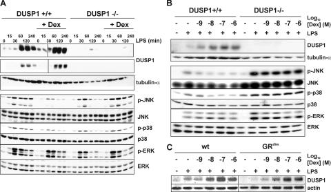
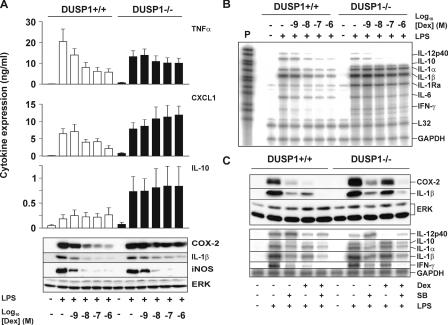
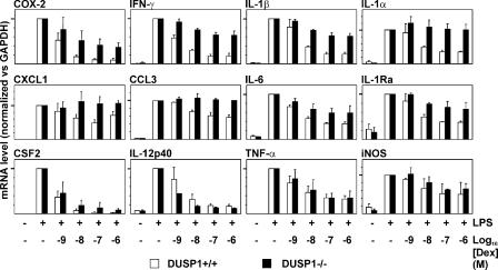
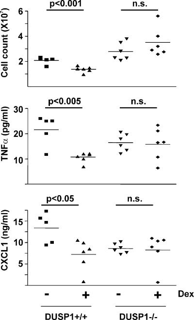
Glucocorticoids (GCs), which are used in the treatment of immune-mediated inflammatory diseases, inhibit the expression of many inflammatory mediators. They can also induce the expression of dual specificity phosphatase 1 (DUSP1; otherwise known as mitogen-activated protein kinase [MAPK] phosphatase 1), which dephosphorylates and inactivates MAPKs. We investigated the role of DUSP1 in the antiinflammatory action of the GC dexamethasone (Dex). Dex-mediated inhibition of c-Jun N-terminal kinase and p38 MAPK was abrogated in DUSP1-/- mouse macrophages. Dex-mediated suppression of several proinflammatory genes (including tumor necrosis factor, cyclooxygenase 2, and interleukin 1alpha and 1beta) was impaired in DUSP1-/- mouse macrophages, whereas other proinflammatory genes were inhibited by Dex in a DUSP1-independent manner. In vivo antiinflammatory effects of Dex on zymosan-induced inflammation were impaired in DUSP1-/- mice. Therefore, the expression of DUSP1 is required for the inhibition of proinflammatory signaling pathways by Dex in mouse macrophages. Furthermore, DUSP1 contributes to the antiinflammatory effects of Dex in vitro and in vivo.


Figures

Figure 1.
Figure 1.
Altered JNK and p38 MAPK signaling in DUSP1−/− BMMs. (A) 106 DUSP1+/+ or DUSP1−/− BMMs were pretreated with vehicle or 100 nM Dex for 4 h and stimulated with LPS for the indicated times. Lysates were blotted for DUSP1 or tubulin-α (top) and total or phosphorylated MAPKs (bottom). Two exposures of the same DUSP1 Western blot are shown to illustrate differences in basal and LPS-induced DUSP1 protein expression. (B) DUSP1+/+ or DUSP1−/− BMMs were pretreated with 1 nM to 1 μM Dex for 4 h and stimulated with LPS for 4 h. In two separate representative experiments, lysates were sequentially blotted for DUSP1 and tubulin-α (top) or for phosphorylated and total MAPKs (bottom). (C) Wild-type and GRdim BMMs were pretreated with 1 nM to 1 μM Dex for 4 h and stimulated with LPS for 4 h. Lysates were blotted for DUSP1 or actin.
Figure 2.
Figure 2.
Antiinflammatory actions of Dex are impaired in DUSP1−/− BMMs. (A) 106 DUSP1+/+ or DUSP1−/− BMMs were pretreated with 1 nM to 1 μM Dex for 4 h and stimulated with LPS for 4 h. Cytokines in supernatants were quantified by ELISA. Graphs indicate means ± SEM (error bars) from at least three independent experiments. Cell lysates were blotted for COX-2, IL-1β, iNOS, and ERK proteins. (B) Cells were treated as in A, and the expression of various mRNAs was examined by RPA. P, undigested probe (10% of input). (C) 106 DUSP1+/+ or DUSP1−/− BMMs were pretreated with 100 nM Dex and/or 10 μM SB202190 and stimulated with LPS for 4 h. Lysates were blotted for Cox-2, IL-1β, or ERK proteins (top), or the expression of various mRNAs was examined by RPA (bottom).
Figure 3.
Figure 3.
Dose-dependent suppression of various inflammatory mediator mRNAs by Dex in DUSP1+/+ and DUSP1−/− BMMs. BMMs were treated as in Fig. 2 B. mRNAs were quantified using qPCR (CSF2, TNF, CXCL1, and COX-2) or RPA. mRNA levels were normalized against GAPDH and expressed as percentages of the level in cells treated with LPS alone. Graphs show mean values ± SEM (error bars) from three to six independent experiments. 100% values are identified by heavy tick marks on the y axes.
Figure 4.
Figure 4.
Impaired antiinflammatory action of Dex in DUSP1−/− mice. Dorsal air pouches were created in DUSP1+/+ and DUSP1−/− mice. Mice were orally dosed with 1 mg/kg Dex or vehicle. After 1 h, zymosan was injected into the air pouches. After a further 4 h, mice were killed, and leukocyte numbers and cytokine concentrations in exudates were quantified. Horizontal bars above graphs represent statistical analysis of differences between control and Dex-treated animals (Student's t test).

References

    1. Newton, R. 2000. Molecular mechanisms of glucocorticoid action: what is important? Thorax. 55:603–613. - PMC - PubMed
    1. Adcock, I.M., and S.J. Lane. 2003. Corticosteroid-insensitive asthma: molecular mechanisms. J. Endocrinol. 178:347–355. - PubMed
    1. Adcock, I.M., and G. Caramori. 2001. Cross-talk between pro-inflammatory transcription factors and glucocorticoids. Immunol. Cell Biol. 79:376–384. - PubMed
    1. Ogawa, S., J. Lozach, C. Benner, G. Pascual, R.K. Tangirala, S. Westin, A. Hoffmann, S. Subramaniam, M. David, M.G. Rosenfeld, and C.K. Glass. 2005. Molecular determinants of crosstalk between nuclear receptors and toll-like receptors. Cell. 122:707–721. - PMC - PubMed
    1. Reichardt, H.M., K.H. Kaestner, J. Tuckermann, O. Kretz, O. Wessely, R. Bock, P. Gass, W. Schmid, P. Herrlich, P. Angel, and G. Schutz. 1998. DNA binding of the glucocorticoid receptor is not essential for survival. Cell. 93:531–541. - PubMed

Publication types

MeSH terms