. 2006 Aug 7;203(8):2021-31.
doi: 10.1084/jem.20060376. Epub 2006 Jul 31.

An inflammatory checkpoint regulates recruitment of graft-versus-host reactive T cells to peripheral tissues

Affiliations

An inflammatory checkpoint regulates recruitment of graft-versus-host reactive T cells to peripheral tissues

Ronjon Chakraverty et al. J Exp Med. .

Abstract

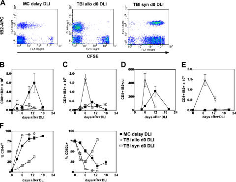
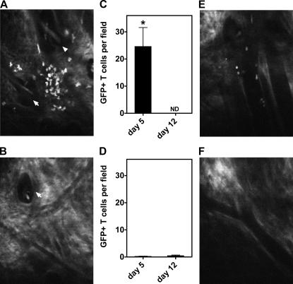
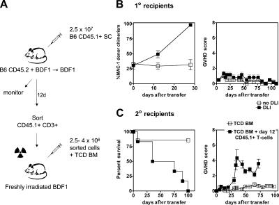
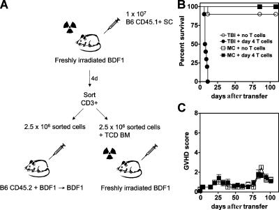
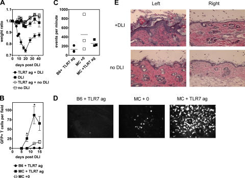
Transfer of T cells to freshly irradiated allogeneic recipients leads to their rapid recruitment to nonlymphoid tissues, where they induce graft-versus-host disease (GVHD). In contrast, when donor T cells are transferred to established mixed chimeras (MCs), GVHD is not induced despite a robust graft-versus-host (GVH) reaction that eliminates normal and malignant host hematopoietic cells. We demonstrate here that donor GVH-reactive T cells transferred to MCs or freshly irradiated mice undergo similar expansion and activation, with similar up-regulation of homing molecules required for entry to nonlymphoid tissues. Using dynamic two-photon in vivo microscopy, we show that these activated T cells do not enter GVHD target tissues in established MCs, contrary to the dogma that activated T cells inevitably traffic to nonlymphoid tissues. Instead, we show that the presence of inflammation within a nonlymphoid tissue is a prerequisite for the trafficking of activated T cells to that site. Our studies help to explain the paradox whereby GVH-reactive T cells can mediate graft-versus-leukemia responses without inducing GVHD in established MCs.


Figures

Figure 1.
Figure 1.
Distribution of antihost CD8+ CTLs between lymphoid and nonlymphoid tissues differs according to host environment. (A) Proliferation of transferred 2C CD8+ T cells as evaluated by CFSE dilution. Dot plots showing CFSE staining (x axis) versus staining with the clonotypic marker 1B2 (y axis) in gated CD8+ T cells derived from recipient spleen on day +6 after transfer to established MCs (MC), freshly irradiated allogeneic recipients (TBI allo), or syngeneic recipients (TBI syn). Note that the CFSE+1B2 population represents polyclonal CFSE-labeled B6 CD8+ T cells that were cotransferred. (B–E) Absolute numbers expressed as mean ± SEM (n = 3 mice per group, except syngeneic mice, which comprised 1–2 mice per group) of antihost 2C transgenic CD8+ CTLs in the spleen (B), lymph nodes (C), peripheral blood (D), and intra-epithelial compartment of the intestine (E) after DLIs on day 0 to freshly irradiated allogeneic (◯) or syngeneic mice (□), or to established MCs (▪). Data shown in A–E are one of three independent experiments performed, which had similar results. Peripheral blood enumeration was not performed in syngeneic control mice. (F) Mean percentage (±SEM) of antihost 2C transgenic CD8+ CTLs in the spleen that were CD44high and CD62L+ after DLIs to MCs (▪) and freshly irradiated allogeneic (◯) or syngeneic mice (□). Data are representative of one out of three independent experiments.
Figure 2.
Figure 2.
Absence of donor T cells in skin after transfer to established MCs. Representative images of skin (A and B, day 5) and mean ± SEM (n = 3 per group) number of GFP+ T cells per field (C and D) derived from two-photon imaging of recipient ear pinnae after transfer to freshly irradiated recipients (A and C) and MCs (B and D) *, P < 0.05 day 5 freshly irradiated mice versus MC. White arrows indicate hair follicles. Representative images of postcapillary venules imaged on day 5 after GFP+ T cell transfer to freshly irradiated recipients (E) or MCs (F). Freshly irradiated mice died before day 12 and therefore could not be assessed at this time point. ND, not done.
Figure 3.
Figure 3.
T cells derived from MCs induce GVHD upon transfer to freshly irradiated recipients. (A) 2.5 × 10⁷ naive B6 CD45.1 splenocytes were transferred to established B6 + BDF1→BDF1 MCs. On day 12, CD45.1+CD3+ T cells were sorted from recipient spleens and 2.5–4 × 10⁶ cells were transferred to secondary irradiated recipients. (B) Transfer of naive donor splenocytes (▪) to MCs induces conversion to full donor chimerism (left) but not GVHD (right) as compared with untreated MCs (□). (C) Survival (left) and clinical GVHD scores (right) after transfer of 4 × 10⁶ day 12 CD45.1+ T cells plus TCD donor BM (▪; n = 6) or TCD BM alone (□; n = 7) to freshly irradiated secondary recipients. Data shows one of two independent experiments with similar results (survival comparison, P = 0.004 day 12 CD45.1+ T cells plus TCD donor BM vs. TCD BM alone).
Figure 4.
Figure 4.
T cells derived from mice developing GVHD are unable to induce GVHD upon transfer to MCs. (A) 107 naive B6 CD45.1 splenocytes plus TCD BM were transferred to freshly irradiated BDF1 recipients. On day 4, CD3+ T cells were sorted from recipient spleens and 2.5 × 10⁶ cells were transferred to secondary irradiated BDF1 mice or to established B6 + BDF1→BDF1 MCs. (B) Survival and (C) clinical GVHD scores after transfer of 2.5 × 10⁶ day 4 T cells plus TCD donor BM (•; n = 10) or TCD BM alone (◯; n = 11) to freshly irradiated secondary recipients, or after transfer of day 4 T cells (▪; n = 11) or nil (□; n = 8) to established MCs. Pooled data are shown from two independent experiments of the same design (P < 0.0001 survival comparison day 4 T cells plus TCD donor BM vs. each of three other groups).
Figure 5.
Figure 5.
Localized TLR-induced inflammation permits access to skin for activated T cells. (A) Mean ± SEM weight ratio of untreated MCs (□; n = 3) or at timed intervals in MCs after transfer of 3 × 10⁷ splenocytes alone (▪; n = 7), 3 × 10⁷ splenocytes plus 50 μg R-848 (given as 12.5 μg by i.p. injections at 3-d intervals from day 0; •; n = 7), or 50 μg R-848 alone (◯; n = 4). (B) Mean ± SEM (n = 3 per group) number of GFP+ T cells per field at time intervals after transfer of 9 × 10⁶ GFP+ T cells to syngeneic control mice treated topically with imiquimod (•), to untreated MCs (□), or to imiquimod-treated MCs (▪). *, P < 0.05 days 8 and 12 treated versus untreated MCs. (C) In vivo flow cytometry was used to examine the frequency of GFP+ T cells in the arteriolar circulation of ear pinnae on day 12 after transfer of 9 × 10⁶ GFP+ T cells to the above groups. (D) Representative images derived from two-photon imaging of recipient ear pinnae on day 12 after transfer of 9 × 10⁶ GFP+ donor T cells to B6 mice treated topically with imiquimod (left), to untreated MCs (middle), or to imiquimod-treated MCs (right). (E) The left flank was treated with imiquimod, and the right flank was left untreated in MCs receiving (top) or not receiving (bottom) 3 × 10⁷ donor splenocytes. Representative images are shown of histology on day 14 after transfer.

Similar articles

Cited by

References

    1. Wysocki, C.A., A. Panoskaltsis-Mortari, B.R. Blazar, and J.S. Serody. 2005. Leukocyte migration and graft-versus-host disease. Blood. 105:4191–4199. - PMC - PubMed
    1. Sprent, J., M. Schaefer, D. Lo, and R. Korngold. 1986. Functions of purified L3T4+ and Lyt-2+ cells in vitro and in vivo. Immunol. Rev. 91:195–218. - PubMed
    1. Shlomchik, W.D., M.S. Couzens, C.B. Tang, J. McNiff, M.E. Robert, J. Liu, M.J. Shlomchik, and S.G. Emerson. 1999. Prevention of graft versus host disease by inactivation of host antigen-presenting cells. Science. 285:412–415. - PubMed
    1. Teshima, T., R. Ordemann, P. Reddy, S. Gagin, C. Liu, K.R. Cooke, and J.L. Ferrara. 2002. Acute graft-versus-host disease does not require alloantigen expression on host epithelium. Nat. Med. 8:575–581. - PubMed
    1. Beilhack, A., S. Schulz, J. Baker, G.F. Beilhack, C.B. Wieland, E.I. Herman, E.M. Baker, Y.A. Cao, C.H. Contag, and R.S. Negrin. 2005. In vivo analyses of early events in acute graft-versus-host disease reveal sequential infiltration of T cell subsets. Blood. 106:1113–1122. - PMC - PubMed

Publication types

Substances