Review
. 2006 Jul 31;174(3):323-7.
doi: 10.1083/jcb.200604035.

Interactions between heparan sulfate and proteins: the concept of specificity

Affiliations
Review

Interactions between heparan sulfate and proteins: the concept of specificity

Johan Kreuger et al. J Cell Biol. .

Abstract

Proteoglycan (PG) coreceptors carry heparan sulfate (HS) chains that mediate interactions with growth factors, morphogens, and receptors. Thus, PGs modulate fundamental processes such as cell survival, division, adhesion, migration, and differentiation. This review summarizes recent biochemical and genetic information that sheds new light on the nature of HS-protein binding. Unexpectedly, many interactions appear to depend more on the overall organization of HS domains than on their fine structure.


Figures

Figure 1.
Figure 1.
Proposed roles of HSPGs in growth factor/morphogen signaling. Locally produced and secreted protein ligands (e.g., growth factors) (1) are captured by HS chains and accumulate at the cell surface (2). Interactions with HS support the generation of protein gradients (3). HS chains promote stable interactions between growth factors and receptors and, thus, modulate the quality of receptor signaling (such as amplitude and kinetics of activation/inactivation; 4). HSPGs may also regulate the turnover of receptors and participate in the internalization of receptor complexes (5). Shedding of HSPG ectodomains (Kreuger et al., 2004) or degradation of HS chains by heparanase (Vlodavsky and Friedmann, 2001) may release HS-bound ligands from the cell surface (6).
Figure 2.
Figure 2.
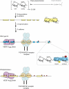
Biosynthesis of HS and molecular phenotypes resulting from deficient HS biosynthetic enzymes. See Esko and Lindahl (2001) and Hacker et al. (2005). HS chains are synthesized while attached to core protein serine residues through a GlcA-Gal-Gal-Xyl linkage region. The linear HS chain is thereafter polymerized through the action of GlcNAc- and GlcA-transferases belonging to the EXT family and further modified by partial N-deacetylation/N-sulfation (N-deacetylase/N-sulfotransferase) to yield NS disaccharide units. Consecutive stretches of such units (NS domains) are hotspots for further modifications: a C5 epimerase converts GlcA to IdoA followed by variable O-sulfation at C-3 and C-6 (red circles) of GlcN and at C-2 (yellow circles) of IdoA (and some GlcA) units. Completed chains may be further edited by endo-6-O-sulfatases (Ai et al., 2003). Protein ligands interact with single NS domains (e.g., FGFs) or with NS domains separated by N-acetylated disaccharide residues (SAS domains; illustrated here for VEGF-A165 [Robinson et al., 2006]; and FGF–HS–FGF receptor complexes). The bottom model depicts a molecular phenotype of C5 epimerase−/− HS that lacks IdoA and IdoA 2-O-sulfation but is more extensively N- and 6-O-sulfated than the corresponding wild-type product. The 2-O-sulfotransferase−/− HS is similar to the C5 epimerase−/− polysaccharide except for the presence of IdoA (Merry et al., 2001). HS from C5 epimerase−/− or 2-O-sulfotransferase−/− cells may still interact more or less efficiently with many protein ligands (see Knockout clues from embryology). Blue boxes, NS domains containing GlcA and/or IdoA; yellow boxes, NS domains containing GlcA but no IdoA.

References

    1. Ai, X., A.T. Do, O. Lozynska, M. Kusche-Gullberg, U. Lindahl, and C.P. Emerson Jr. 2003. QSulf1 remodels the 6-O sulfation states of cell surface heparan sulfate proteoglycans to promote Wnt signaling. J. Cell Biol. 162:341–351. - PMC - PubMed
    1. Allen, B.L., and A.C. Rapraeger. 2003. Spatial and temporal expression of heparan sulfate in mouse development regulates FGF and FGF receptor assembly. J. Cell Biol. 163:637–648. - PMC - PubMed
    1. Ashikari-Hada, S., H. Habuchi, Y. Kariya, N. Itoh, A.H. Reddi, and K. Kimata. 2004. Characterization of growth factor-binding structures in heparin/heparan sulfate using an octasaccharide library. J. Biol. Chem. 279:12346–12354. - PubMed
    1. Ashikari-Hada, S., H. Habuchi, Y. Kariya, and K. Kimata. 2005. Heparin regulates vascular endothelial growth factor165-dependent mitogenic activity, tube formation, and its receptor phosphorylation of human endothelial cells. Comparison of the effects of heparin and modified heparins. J. Biol. Chem. 280:31508–31515. - PubMed
    1. Bernfield, M., R. Kokenyesi, M. Kato, M.T. Hinkes, J. Spring, R.L. Gallo, and E.J. Lose. 1992. Biology of the syndecans: a family of transmembrane heparan sulfate proteoglycans. Annu. Rev. Cell Biol. 8:365–393. - PubMed

Publication types