. 2006 Jul 31;174(3):459-71.
doi: 10.1083/jcb.200508170.

Tetanus toxin is internalized by a sequential clathrin-dependent mechanism initiated within lipid microdomains and independent of epsin1

Affiliations

Tetanus toxin is internalized by a sequential clathrin-dependent mechanism initiated within lipid microdomains and independent of epsin1

Katrin Deinhardt et al. J Cell Biol. .

Abstract

Ligand-receptor complexes are internalized by a variety of endocytic mechanisms. Some are initiated within clathrin-coated membranes, whereas others involve lipid microdomains of the plasma membrane. In neurons, where alternative targeting to short- or long-range trafficking routes underpins the differential processing of synaptic vesicle components and neurotrophin receptors, the mechanism giving access to the axonal retrograde pathway remains unknown. To investigate this sorting process, we examined the internalization of a tetanus neurotoxin fragment (TeNT HC), which shares axonal carriers with neurotrophins and their receptors. Previous studies have shown that the TeNT HC receptor, which comprises polysialogangliosides, resides in lipid microdomains. We demonstrate that TeNT HC internalization also relies on a specialized clathrin-mediated pathway, which is independent of synaptic vesicle recycling. Moreover, unlike transferrin uptake, this AP-2-dependent process is independent of epsin1. These findings identify a pathway for TeNT, beginning with the binding to a lipid raft component (GD1b) and followed by dissociation from GD1b as the toxin internalizes via a clathrin-mediated mechanism using a specific subset of adaptor proteins.


Figures
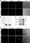
Figure 1.
Figure 1.
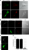
TeNT HC internalization is independent of SV exocytosis and recycling. (A) MNs were incubated with 20 nM Alexa Fluor 555–TeNT HC for 30 min at 37°C, either under resting conditions (a–d) or after stimulation of SV exo/endocytosis by adding 60 mM KCl to the medium just before TeNT HC addition (e–h), fixed, and stained for VAMP-2. Only very limited colocalization of TeNT HC and VAMP-2 under resting or stimulated conditions, which were quantified in B, was found. Error bars represent the SEM. (C and D) MNs were incubated with 15 nM BoNT/A and 2 nM BoNT/D for 22 h at 37°C to cleave SNAP-25 and VAMP-2. Untreated cells were processed in parallel for comparison. (C) Cells were scraped and analyzed by Western blotting using antibodies raised against the cleaved fragments of SNAP-25 and VAMP-2, as well as actin, as a loading control. (D) 20 nM b-TeNT HC was added to MNs for 30 min at 37°C. MNs were shifted to ice, treated with MESNA before fixation, and stained for VAMP-2 (a and e), SNAP-25 (b and f), and biotin (c and g). Pretreatment with BoNTs did not affect TeNT HC internalization. DIC, differential interference contrast. Bars: (A) 5 μm; (D) 10 μm.
Figure 2.
Figure 2.
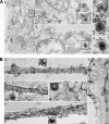
Dynamin is required for TeNT HC uptake into MNs. (A) MNs were incubated with 50 μM of the cell-permeable peptide P4 (top) or its scrambled analogue P4S (bottom) for 2 h before addition of 20 nM Alexa Fluor 488–TeNT HC and 0.2 mg/ml tetramethylrhodamine-dextran for additional 45 min. Images show confocal sections through the cell body. (B–D) MNs were microinjected with a plasmid encoding Myc-dynamin2K44A. (B) After 25 h of expression, cells were incubated with 20 nM b-TeNT HC and 0.2 mg/ml tetramethylrhodamine-dextran for 30 min at 37°C, treated with MESNA, fixed, stained for internal biotin and with an anti-Myc antibody to detect the tagged dynamin2K44A, and imaged. (top) A control MN; (bottom) a microinjected cell. (C) Expression of dynamin2K44A does not block the surface binding of TeNT HC to MNs. 25 h after microinjection of dynamin2K44A, MNs were incubated with Alexa Fluor 647–TeNT HC for 30 min at 37°C, fixed, and stained with an anti-Myc antibody. A confocal section of an expressing cell is shown. (D) Quantitative analysis of the effect of dynamin2K44A overexpression on TeNT HC internalization. Noninjected MNs or cells microinjected with a GFP-encoding plasmid were taken as controls. Numbers in parenthesis indicate the number of MNs imaged per condition. DIC, differential interference contrast. Bars: (A) 5 μm; (B and C) 10 μm.
Figure 3.
Figure 3.
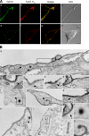
TeNT HC enters clathrin-coated structures in MNs. (A and B) MNs were microinjected with a plasmid encoding GFP-CLC. After overnight expression, cells were incubated with 30 nM Alexa Fluor 555–TeNT HC, washed, and imaged. (A) GFP-CLC and TeNT HC dual-positive structures are visible in an axon (arrows). (B) Confocal section of the bottom plane of a MN soma. A partial overlap between GFP-CLC–positive and TeNT HC–positive structures is observed (inset). (C) MNs were incubated with HRP–TeNT HC for 45 min on ice, and then chased for 45 min at 12 (a and b) or 18°C (c). Cells were fixed and incubated with DAB/H2O2 to label HRP–TeNT HC. The electron-dense DAB reaction product was often associated with coated pits and budding vesicles, which were visible at higher magnification (b' and c'). Bars: (A) 2 μm; (B) 5 μm; (C) 0.2 μm.
Figure 4.
Figure 4.
Colocalization of HRP–TeNT HC and clathrin at EM level. (A) HRP–TeNT HC–containing structures are labeled with anti-CHC. Cells were loaded with HRP–TeNT HC at 12°C for 45 min before HRP cross-linking with DAB/H2O2. MNs were permeabilized with digitonin before labeling with an anti-CHC antibody and incubated with a rabbit anti–mouse bridging antibody, and then with 10 nm immunogold. (B) Whole-mount transmission EM showing distribution of clathrin in vesicles loaded with HRP–TeNT HC. (a–d) MNs were incubated with HRP–TeNT HC at 12°C for 45 min, cross-linked with DAB/H2O2, extracted with Triton X-100, and then treated with an anti-CHC antibody followed by a bridging antibody and 10 nm immunogold. HRP–TeNT HC is found in CCPs and CCVs in axons (a, b, d) and soma (c). Control without the bridging antibody showed negligible labeling (e, arrows). Boxed areas show enlarged examples of TENT HC–positive endocytic structures. Bars, 0.2 μm.
Figure 5.
Figure 5.
GD1b and TeNT HC undergo independent sorting on the plasma membrane. MNs were probed with TeNT HC and an anti-GD1b antibody and analyzed at light and EM levels. (A) Cells were incubated with the antibody MOG-1 (2 μg/ml) and 20 nM Alexa Fluor 555–TeNT HC for 30 min on ice, followed by 5 min at 22°C, and then were washed, fixed, and imaged. A good overlap of the two signals could be seen both on neurites (a–d) and soma (e–h). DIC, differential interference contrast. (B) EM analysis. MNs were incubated on ice with 60 nM HRP–TeNT HC and 10 μg/ml MOG-1, followed by 10 nm immunogold. After washing, samples were shifted to 12°C for 45 min (a–h) or 37°C for 15 min (i). MNs were fixed and incubated with DAB/H2O2 to label HRP–TeNT HC. The anti-GD1b gold-labeled antibody is excluded from endocytosed HRP–TeNT HC vesicles (asterisks), clathrin lattices (arrows), and invaginated coated pits (arrowheads). Bars, 0.2 μm.
Figure 6.
Figure 6.
Internalization of TeNT HC is dependent on a functional clathrin machinery. 25 h after microinjection of the AP-2 μ2T156A and tTA plasmids (d–f), or 26 h after microinjection of a plasmid encoding AP180-C (g–i), MNs were incubated with 20 nM b-TeNT HC and 10 ng/ml Alexa Fluor 555–CTB for 30 min, MESNA-treated, fixed, stained for internal biotin and the epitope tags of the microinjected constructs, and imaged. (a–c) Uninjected MNs were imaged as a control. Bar, 10 μm.
Figure 7.
Figure 7.
TeNT HC endocytosis is independent of epsin1. MNs were microinjected with a plasmid encoding epsin1R63L H73L. (A) After 26 h, cells were incubated with 20 nM b-TeNT HC and 20 μg/ml Alexa Fluor 594–transferrin or 10 ng/ml Alexa Fluor 555–CTB for 30 min at 37°C and then shifted on ice for MESNA treatment. MNs were fixed and stained for internal biotin and with an anti-Myc antibody before imaging. Control cells readily internalized both transferrin and TeNT HC (a–c), whereas transferrin, but not TeNT HC uptake was blocked in microinjected cells (d, f, and g). CTB internalization was also not affected in epsin1R63L H73L-expressing cells (i). (B) 26 h after comicroinjection with plasmids encoding epsin1R63L H73L and HRP-KDEL, MNs were incubated with HRP–TeNT HC for 30 min at 37°C. Cells were then treated with DAB/H2O2 and analyzed by EM. Transfection was confirmed by the characteristic DAB staining of the tubular ER (asterisks). The DAB reaction product generated by HRP–TeNT HC was found in various endosomes, multivesicular body–like structures (arrows), and in CCVs (arrowheads and insets). (C) Quantification of the effects of the overexpression of dominant-negative mutants on TeNT HC endocytosis as determined in Fig. 5 and Fig. 6 A. Numbers in parenthesis indicate the number of MNs observed per condition. Bars: (A) 10 μm; (B) 0.2 μm.
Figure 8.
Figure 8.
Internalization pathways in MNs. Transferrin uptake is mediated by a classical clathrin-dependent internalization route occurring in soma and dendrites. SV exo/endocytosis accounts for the majority of endocytic events at the presynaptic terminal and may involve multiple clathrin-dependent steps. In contrast, CTB, which binds to GM1 clustered in lipid rafts, is internalized by a clathrin-independent, dynamin-dependent mechanism in MNs. TeNT HC exploits a pathway requiring lipid rafts and the clathrin machinery, which is distinct from aforementioned routes of internalization. At the NMJ, TeNT HC binds to a lipid–protein receptor complex containing the ganglioside GD1b. TeNT HC is then laterally sorted into CCPs and, during this sorting event, GD1b is excluded from the toxin receptor complex. Internalization of TeNT HC is dependent on dynamin, AP-2, and AP180, but does not require epsin1. Once internalized, TeNT HC is targeted to a stationary early sorting compartment (Lakadamyali et al., 2006), to which other endocytic routes may converge. This early sorting compartment is functionally coupled to the axonal retrograde transport pathway.

Similar articles

Cited by

References

    1. Abrami, L., S. Liu, P. Cosson, S.H. Leppla, and F.G. van der Goot. 2003. Anthrax toxin triggers endocytosis of its receptor via a lipid raft-mediated clathrin-dependent process. J. Cell Biol. 160:321–328. - PMC - PubMed
    1. Aquino, D.A., M.A. Bisby, and R.W. Ledeen. 1985. Retrograde axonal transport of gangliosides and glycoproteins in the motoneurons of rat sciatic nerve. J. Neurochem. 45:1262–1267. - PubMed
    1. Blanpied, T.A., D.B. Scott, and M.D. Ehlers. 2002. Dynamics and regulation of clathrin coats at specialized endocytic zones of dendrites and spines. Neuron. 36:435–449. - PubMed
    1. Boffey, J., M. Odaka, D. Nicoll, E.R. Wagner, K. Townson, T. Bowes, J. Conner, K. Furukawa, and H.J. Willison. 2005. Characterisation of the immunoglobulin variable region gene usage encoding the murine anti-ganglioside antibody repertoire. J. Neuroimmunol. 165:92–103. - PubMed
    1. Bohnert, S., and G. Schiavo. 2005. Tetanus toxin is transported in a novel neuronal compartment characterized by a specialized pH regulation. J. Biol. Chem. 280:42336–42344. - PubMed

Publication types

MeSH terms