. 2006 Aug 8;103(32):11934-9.
doi: 10.1073/pnas.0510306103. Epub 2006 Jul 31.

Stimulation of phosphatidylinositol kinase type I-mediated phosphatidylinositol (4,5)-bisphosphate synthesis by AP-2mu-cargo complexes

Affiliations

Stimulation of phosphatidylinositol kinase type I-mediated phosphatidylinositol (4,5)-bisphosphate synthesis by AP-2mu-cargo complexes

Michael Krauss et al. Proc Natl Acad Sci U S A. .

Abstract

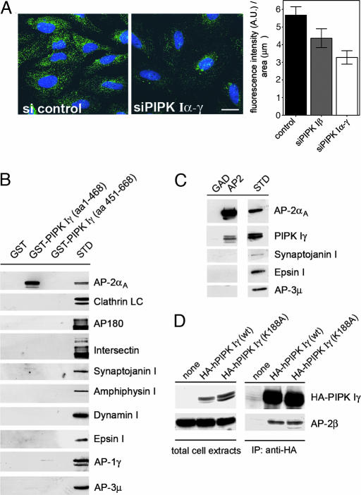
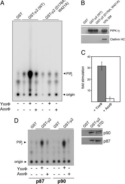
Phosphatidylinositol (4,5)-bisphosphate [PI(4,5)P(2)] is an important factor for a variety of cellular functions ranging from cell signaling to actin cytoskeletal dynamics and endocytic membrane traffic. Here, we have identified the clathrin adaptor complex AP-2 as a regulator of phosphatidylinositol 4-phosphate 5-kinase (PIPK)-mediated PI(4,5)P(2) synthesis. AP-2 directly interacts with the kinase core domain of type I PIPK isozymes via its mu2-subunit in vitro and in native protein extracts. Endocytic cargo protein binding to mu2 leads to a potent stimulation of PIPK activity. These data thus identify a positive feedback loop consisting of endocytic cargo proteins, AP-2mu, and PIPK type I which may provide a specific pool of PI(4,5)P(2) dedicated to clathrin/AP-2-dependent receptor internalization.


Conflict of interest statement

Conflict of interest statement: No conflicts declared.

Figures

Fig. 1.
Fig. 1.
Cargo-containing clathrin/AP-2-coated pits form along PIPK-positive plasma membrane areas. (A) COS7 fibroblasts expressing HA-PIPK type Iα (blue) were incubated with Texas red-labeled EGF at 8°C for 60 min, washed, fixed, and immunostained for AP-2α (green). (Scale bar, 10 μm.) (B) Boxed area in A. The majority of EGF-positive AP-2-coated pits emanates from PIPK Iγ-containing membrane patches. Arrows denote spots of colocalization of AP-2 with both EGF (Lower Left), and PIPK Iγ (Lower Right). (Scale bar, 10 μm.) (C and D) Same as in A and B upon expression of HA-PIPK Iγ (K188A).
Fig. 2.
Fig. 2.
PIPK Iγ directly interacts with AP-2μ. (A) RNAi-mediated knockdown of PIPK I attenuates AP-2-coated pit staining in HeLa cells. (Left and Center) Fixed cells were analyzed for AP-2α-staining. Total fluorescence intensity was quantified and normalized to the area covered by the cells. (Right) Data are depicted as mean (±SD). (Scale bar, 20 μm.) (B) The kinase core domain of PIPK Iγ interacts with AP-2 but not AP-1 or AP-3. Immunoblot analysis of material affinity-purified using GST, GST-tagged PIPK Iγ kinase (1–468), or tail domains (451–668) is shown. γ- and μ3-Adaptins are subunits of AP-1 or AP-3 complexes, respectively. STD, 2.5% of the total amount of Triton X-100-extracted rat brain lysates added to the assay. (C) PIPK Iγ coimmunoprecipitates with AP-2 from rat brain lysates. Detergent-lysed rat brain extracts were subjected to immunoprecipation using monoclonal antibodies against AP-2α. Samples were washed and analyzed by immunoblotting for the proteins indicated. STD, 5% of the total amount of Triton X-100-extracted rat brain lysates added to the assay. (D) AP-2 coimmunoprecipitates with HA-PIPK Iγ or a kinase-dead mutant (K188A) from transfected fibroblasts. Detergent extracts of transfected HEK293 cells expressing wild-type PIPK Iγ or a kinase-inactive mutant (K188A) were subjected to immunoprecipitation using anti-HA antibodies. Aliquots of the total cell extracts and the immunoprecipitated material were analyzed by immunoblotting with antibodies against AP-2β or HA-PIPK Iγ. Cells not expressing HA-PIPK Iγ (none) were taken as a control.
Fig. 3.
Fig. 3.
Interaction of type I PIP kinases with AP-2. (A) PIPK Iγ binds to AP-2 via its μ2-subunit. GST or GST-PIPK Iγ (1–468) were incubated with [35S]-radiolabeled AP-2 subunits synthesized individually by coupled transcription/translation in vitro. After extensive washes, bound proteins were eluted and analyzed by SDS/PAGE and autoradiography. STD, 20% of the total amount of radiolabeled protein added to the assay. (B) AP-2μ associates with all three isoforms of PIPK type I from a detergent extract of transfected fibroblasts. COS7 cells were transfected with HA-tagged PIPK I enzymes; a detergent extract was affinity-purified by using GST or GST-C-μ2, and the affinity-purified material was analyzed by immunoblotting using antibodies recognizing clathrin heavy chain (HC) or HA. (C) Soluble C-μ2-EGFP coimmunoprecipitates with HA-tagged PIPK type I isoforms from transfected fibroblasts. Detergent extracts of transfected Cos7 cells coexpressing soluble C-μ2 (residues 164–435)-EGFP together with PIPK I (α, β, or γ) were subjected to immunoprecipitation using anti-HA-antibodies. Aliquots of the immunoprecipitated material or the supernatants were analyzed with antibodies against EGFP, HA, or AP-1γ. (D) Binding of His6-C-μ2 to the PIPK Iγ kinase core domain determined by surface plasmon resonance. GST-PIPK Iγ (amino acid 1–468) or GST were immobilized on a sensor chip, and His6-C-μ2 was injected at concentrations between 62 nM and 1 μM (lower to upper curves). KD was calculated from the kinetic rate constants derived from the sensograms.
Fig. 4.
Fig. 4.
PIPK Iγ preferentially associates with AP-2 bound to membranes or endocytic receptor sorting signals. (A) AP-2-PIPK Iγ complexes can be isolated preferentially from membrane fractions. Detergent extracts of HEK293 cells inducibly expressing PIPK Iγ were subjected to immunoprecipitation using anti-HA affinity tag antibodies. Aliquots of the material immunoprecipitated from total cell lysates or cytosolic or membrane fractions were analyzed by immunoblotting with antibodies against AP-2α, talin, or HA-PIPK Iγ. STD, detergent extracted rat brain lysates (15 μg of total protein). (B) PIPK Iγ associates with AP-2 bound to the cytoplasmic tail of the EGFR. GST-stonin 1(1–33), the GST-tagged cytoplasmic tail of the EGF receptor (EGFR), or GST were used for affinity purification from detergent-extracted rat brain lysates. Samples were analyzed by SDS/PAGE and immunoblotting for AP-2μ or PIPK Iγ. STD, 5% of the total amount of Triton X-100-extracted rat brain lysates added to the assay. (C) AP-2 coimmunoprecipitates with both EGFR and PIPK Iγ. Detergent extracts of Cos7 cells that had been mock-treated or transfected with plasmids encoding HA-PIPK Iγ and the EGFRs were subjected to immunoprecipitation using antibodies against AP-2α. Aliquots of the supernatant or pellet fractions were analyzed by immunoblotting for AP-2α, EGFR, or HA-PIPK Iγ.
Fig. 5.
Fig. 5.
AP-2μ-tyrosine motif sorting signal complexes activate the PI(4,5)P2-synthesizing activity of PIPK Iγ. (AC) AP-2μ bound to tyrosine-based endocytic sorting signals stimulates PIPK Iγ-mediated PI(4,5)P2 synthesis. PIPK Iγ was affinity-purified from detergent lysates of HEK293 flip-in cells using GST-μ2 or a Yxxø-motif binding-defective mutant [GST-μ2 (D176A,W421A)]. An aliquot of this material was subjected to SDS/PAGE and immunoblotting. (B) Radiolabeled PI(4,5)P2 synthesized in the presence or absence of peptides together with [32P]ATP and PI(4)P was analyzed by TLC and quantitative phosphorimage analysis. Representative data are shown in A. Quantifications are depicted in C. Data (mean stimulation ± SD) derived from three independent experiments were normalized to the amount of PI(4,5)P2 synthesized by GST-μ2-associated PIPK Iγ in the absence of peptide. (D) PI(4,5)P2 synthesis by the p87 and p90 isoforms of PIPK Iγ is stimulated equally well by AP-2μ–Yxxø complexes (Left). The experiment was essentially done as described in A. Similar amounts of p87 or p90 were found to be associated with GST-μ2 (Right). STD, 5% of the total amount of Triton X-100-extracted HEK293 cell lysates added to the assay.

References

    1. Martin T. F. Curr. Opin. Cell Biol. 2001;13:493–499. - PubMed
    1. Yin H. L., Janmey P. A. Annu. Rev. Physiol. 2003;65:761–789. - PubMed
    1. Weernink P. A., Meletiadis K., Hommeltenberg S., Hinz M., Ishihara H., Schmidt M., Jakobs K. H. J. Biol. Chem. 2004;279:7840–7849. - PubMed
    1. Jost M., Simpson F., Kavran J. M., Lemmon M. A., Schmid S. L. Curr. Biol. 1998;8:1399–1402. - PubMed
    1. Donaldson J. G. J. Biol. Chem. 2003;278:41573–41576. - PubMed

Publication types

Substances