

Topical vitamin D3 and low-calcemic analogs induce thymic stromal lymphopoietin in mouse keratinocytes and trigger an atopic dermatitis
- PMID: 16880407
- PMCID: PMC1544239
- DOI: 10.1073/pnas.0604575103
Topical vitamin D3 and low-calcemic analogs induce thymic stromal lymphopoietin in mouse keratinocytes and trigger an atopic dermatitis
Abstract
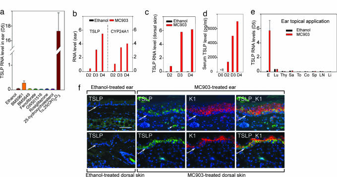
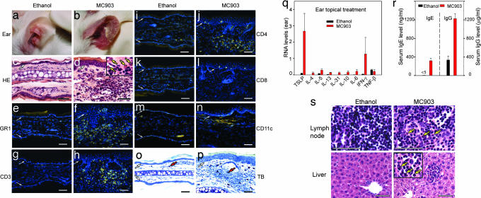
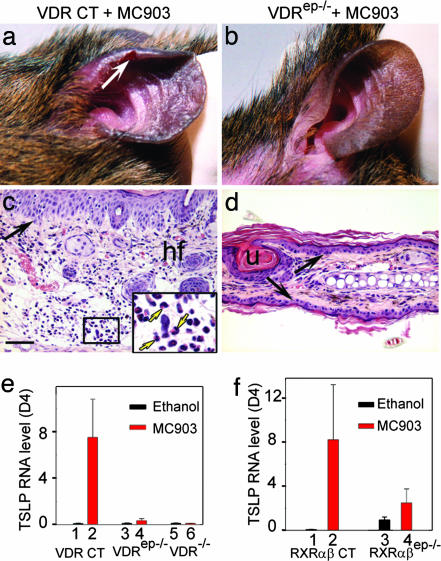
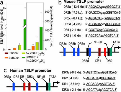
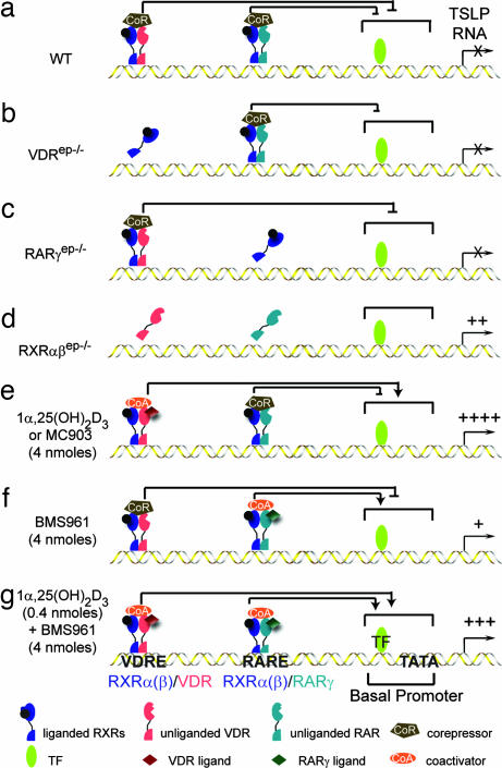
We have demonstrated that cytokine thymic stromal lymphopoietin (TSLP), whose expression is rapidly induced upon keratinocyte-selective ablation of retinoid X receptors (RXRs) -alpha and -beta in the mouse (RXRalphabeta(ep-/-) mice), plays a key role in initiating a skin and systemic atopic dermatitis-like phenotype. We show here that topical application of the physiologically active ligand [1alpha,25-(OH)(2)D(3); calcitriol] of the vitamin D receptor, or of its low-calcemic analog MC903 (calcipotriol; Dovonex), induces TSLP expression in epidermal keratinocytes, which results in an atopic dermatitis-like syndrome mimicking that seen in RXRalphabeta(ep-/-) mutants and transgenic mice overexpressing TSLP in keratinocytes. Furthermore, topical application of retinoic acid receptor RARgamma-selective agonist BMS961 also induces TSLP expression either on its own or synergistically with 1alpha,25-(OH)(2)D(3). Our data demonstrate that RXR/vitamin D receptor and RXR/retinoic acid receptor-gamma heterodimers and their ligands cell-autonomously control the expression of TSLP in epidermal keratinocytes of the mouse. We propose molecular mechanisms through which vitamin D3 and retinoic acid signalings could be involved in the pathogenesis of atopic diseases.
Conflict of interest statement
Conflict of interest statement: No conflicts declared.
Figures
Similar articles
-
TSLP expression in the skin is mediated via RARγ-RXR pathways.Immunobiology. 2016 Feb;221(2):161-5. doi: 10.1016/j.imbio.2015.09.013. Epub 2015 Sep 10. Immunobiology. 2016. PMID: 26531761
-
Induction of thymic stromal lymphopoietin expression in keratinocytes is necessary for generating an atopic dermatitis upon application of the active vitamin D3 analogue MC903 on mouse skin.J Invest Dermatol. 2009 Feb;129(2):498-502. doi: 10.1038/jid.2008.232. Epub 2008 Jul 24. J Invest Dermatol. 2009. PMID: 18650845 No abstract available.
-
Similarities and differences in the transcriptional control of expression of the mouse TSLP gene in skin epidermis and intestinal epithelium.Proc Natl Acad Sci U S A. 2017 Feb 7;114(6):E951-E960. doi: 10.1073/pnas.1620697114. Epub 2017 Jan 23. Proc Natl Acad Sci U S A. 2017. PMID: 28115699 Free PMC article.
-
TSLP expression: cellular sources, triggers, and regulatory mechanisms.Allergol Int. 2012 Mar;61(1):3-17. doi: 10.2332/allergolint.11-RAI-0395. Epub 2012 Jan 25. Allergol Int. 2012. PMID: 22270071 Review.
-
Analysis of the mechanism for the development of allergic skin inflammation and the application for its treatment:keratinocytes in atopic dermatitis - their pathogenic involvement.J Pharmacol Sci. 2009 Jul;110(3):260-4. doi: 10.1254/jphs.09r06fm. J Pharmacol Sci. 2009. PMID: 19609063 Review.
Cited by
-
Elevated epidermal thymic stromal lymphopoietin levels establish an antitumor environment in the skin.Cancer Cell. 2012 Oct 16;22(4):494-505. doi: 10.1016/j.ccr.2012.08.017. Cancer Cell. 2012. PMID: 23079659 Free PMC article.
-
Essential contribution of IRF3 to intestinal homeostasis and microbiota-mediated Tslp gene induction.Proc Natl Acad Sci U S A. 2012 Dec 18;109(51):21016-21. doi: 10.1073/pnas.1219482110. Epub 2012 Dec 3. Proc Natl Acad Sci U S A. 2012. PMID: 23213237 Free PMC article.
-
Keratinocyte-derived cytokine TSLP promotes growth and metastasis of melanoma by regulating the tumor-associated immune microenvironment.JCI Insight. 2022 Nov 8;7(21):e161438. doi: 10.1172/jci.insight.161438. JCI Insight. 2022. PMID: 36107619 Free PMC article.
-
TSLP is differentially regulated by vitamin D3 and cytokines in human skin.Immun Inflamm Dis. 2015 Mar;3(1):32-43. doi: 10.1002/iid3.48. Epub 2015 Feb 16. Immun Inflamm Dis. 2015. PMID: 25866638 Free PMC article.
-
Genomic control of inflammation in experimental atopic dermatitis.Sci Rep. 2022 Nov 7;12(1):18891. doi: 10.1038/s41598-022-23042-x. Sci Rep. 2022. PMID: 36344555 Free PMC article.
References
-
- Laudet V., Gronemeyer H. The Nuclear Receptor: Factsbook. San Diego: Academic; 2002.
-
- Soumelis V., Reche P. A., Kanzler H., Yuan W., Edward G., Homey B., Gilliet M., Ho S., Antonenko S., Lauerma A., et al. Nat. Immunol. 2002;3:673–680. - PubMed
Publication types
MeSH terms
Substances
LinkOut - more resources
Full Text Sources
Other Literature Sources
Molecular Biology Databases
