. 2006 Aug;26(16):6024-36.
doi: 10.1128/MCB.02354-05.

Wnt-5a/Ca2+-induced NFAT activity is counteracted by Wnt-5a/Yes-Cdc42-casein kinase 1alpha signaling in human mammary epithelial cells

Affiliations

Wnt-5a/Ca2+-induced NFAT activity is counteracted by Wnt-5a/Yes-Cdc42-casein kinase 1alpha signaling in human mammary epithelial cells

Janna Dejmek et al. Mol Cell Biol. 2006 Aug.

Abstract

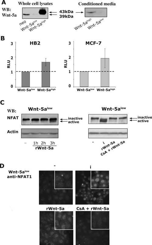
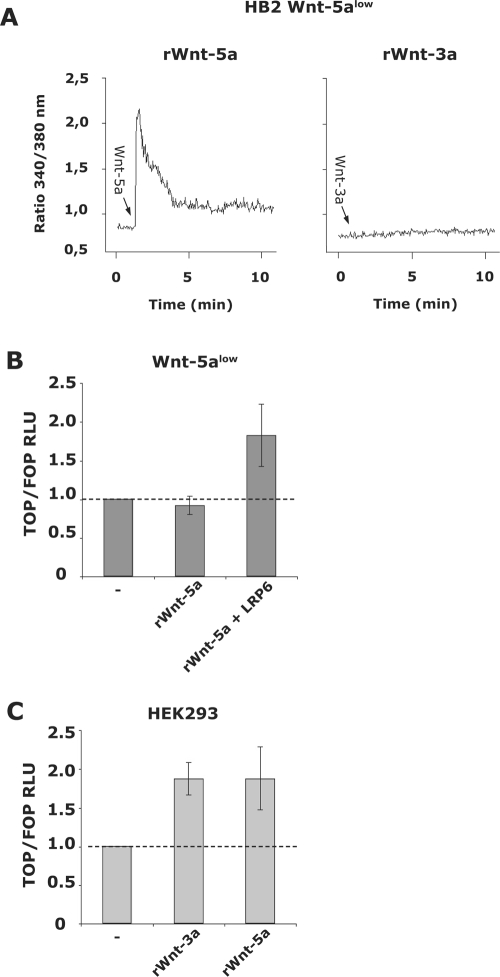
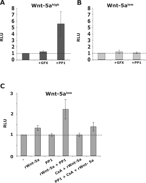
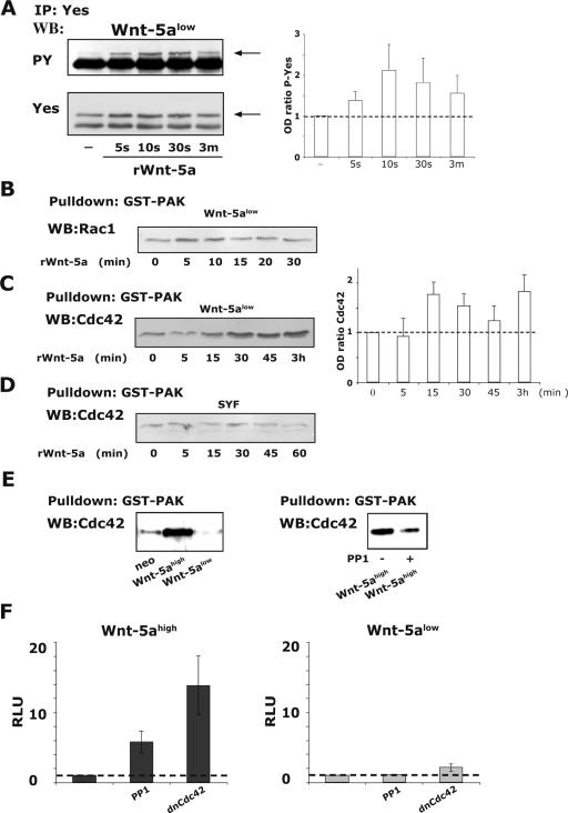
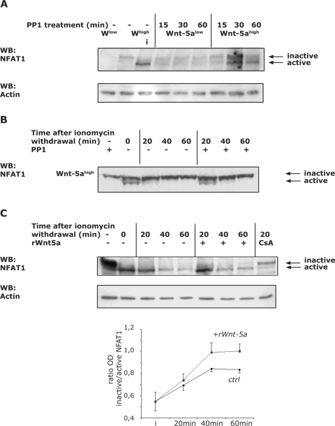
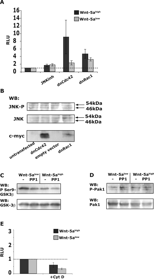
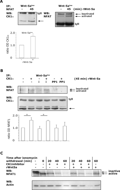
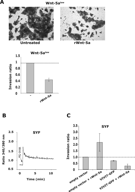
Wnt-5a has been shown to influence the metastatic behavior of human breast cancer cells, and the loss of Wnt-5a expression is associated with metastatic disease. We show here that NFAT1, a transcription factor connected with breast cancer metastasis, is activated by Wnt-5a through a Ca2+ signaling pathway in human breast epithelial cells. This activation was simultaneously counteracted by a Wnt-5a-induced Yes/Cdc42 signaling pathway. The observation that inhibition of the Wnt-5a/Yes/Cdc42 signal prolonged the duration of ionomycin-induced NFAT1 activation revealed the general importance of this pathway. The Wnt-5a-induced inhibition of NFAT1 did not require glycogen synthase kinase 3beta, JNK, or Pak1 activity or modulation of the cytoskeleton. Instead, we observed that Wnt-5a induced a complex formation of NFAT1/casein kinase 1alpha, even upon treatment with ionomycin, which was blocked upon inhibition of the Wnt-5a/Yes/Cdc42 signaling pathway. Our results explain why Wnt-5a/Ca2+-induced NFAT activity is hard to detect and suggest a novel mechanism by which Wnt-5a can suppress tumor-specific, agonist-induced NFAT activity and thus the metastatic behavior of breast cancer cells.


Figures

FIG. 1.
FIG. 1.
(A) Western blot analysis of Wnt-5a expression levels in stably transfected HB2 human breast epithelial cells (left) and concentrated conditioned media (right). To ensure equal loading, protein measurements were done using a Coomassie protein assay reagent (Pierce, Rockford, IL). The blot shown is representative of several separate experiments. In the text, the studied clones are referred to as HB2 neo (empty vector), Wnt-5alow (Wnt-5a antisense), and Wnt-5ahigh (+Wnt-5a HA) cells. (B) Levels of NFAT activation in Wnt-5alow and Wnt-5ahigh HB2 (left) and MCF-7 (right) cells. The activation level of NFAT was measured by using luciferase reporter vectors containing NFAT sites (NFAT-pGL2; AP1 independent). Each transfection was done with approximately 106 cells, 1 μg of reporter gene construct, and 0.2 μg of CMV-controlled Renilla reporter gene. The cells were transfected overnight, after which conditioned medium from either Wnt-5alow or Wnt-5ahigh cells was added. The obtained luciferase activity (measured in relative luciferase units [RLU]) was normalized against the activity of the cotransfected Renilla reporter gene and the TATA-background in the two different cell clones. The statistically significant difference was analyzed by using the Student t test (P < 0.05). (C) Analysis of phosphorylation status of NFAT1 by SDS-6% PAGE upon treatment of Wnt-5alow cells with 1.6 μg of recombinant mouse Wnt-5a (rWnt-5a)/ml for the times indicated. Inactive NFAT is phosphorylated and migrates at 135 kDa, whereas active NFAT1 is dephosphorylated and migrates at 120 kDa. Ionomycin (i) is used as a positive control and CS (CsA) inhibits NFAT activity. (D) Nuclear localization of NFAT1 upon treatment of Wnt-5alow cells with 1.6 μg of recombinant Wnt-5a/ml for 1 h. Ionomycin (i) is used as a positive control, and CS inhibits NFAT activity.
FIG. 2.
FIG. 2.
(A) rWnt-5a (0.8 μg/ml; left) and rWnt-3a (100 ng/ml; right) induced Ca2+ signal in Wnt-5alow HB2 cells. (B) rWnt-5a (0.8 μg/ml; 24 h) induced canonical Wnt signaling as measured by luciferase assays using TOPflash and FOPflash reporters. The obtained luciferase activity (measured in RLU) was normalized against the activity of the cotransfected Renilla reporter gene. Wnt-5a only induces canonical signaling in human breast epithelial cells in the presence of the coreceptor LRP6. (C) rWnt-3a (100 ng/ml; 24 h) and rWnt-5a (0.8 μg/ml; 24 h) induced canonical Wnt signaling in HEK293 cells, as measured by luciferase assays using TOPflash and FOPflash reporters cotransfected with Renilla.
FIG. 3.
FIG. 3.
(A and B) Wnt-5ahigh (A; dark) and Wnt-5alow (B; light) cells were treated for 36 h with GFX (2 μM) or PP1 (10 μM) after which NFAT activation was evaluated as described above (GFX and PP1 added to the conditioned media). The basal levels of NFAT activation were set to 1 for each cell clone. The data are expressed as means ± the standard error of the mean (SEM) of 6 to 12 separate experiments. (C) Effect of rWnt-5a on NFAT activation alone or in combination with PP1 or CS in Wnt-5alow cells as measured by Luciferase reporter assays. Wnt-5alow cells were transfected and treated for 36 h with 0.8 μg of rWnt-5a/ml, 10 μM PP1, or 100 ng of CS/ml. The data are expressed as means ± the SEM of six experiments.
FIG. 4.
FIG. 4.
(A) rWnt-5a induced activation of the Src-kinase Yes in Wnt-5alow cells as measured by determining the phosphotyrosine (PY) levels (arrow; top) of immunoprecipitated Yes. The membranes were reprobed for total Yes (arrow; bottom). The ratio of the optical density (OD) from several experiments is expressed as means ± the SEM (right). (B) rWnt-5a-induced Rac1 activation in Wnt-5alow cells, as measured by GST-pulldown assays of active Rac1. (C) rWnt-5a-induced Cdc42 activation in Wnt-5alow cells, as measured by GST-pulldown assays of active Cdc42. The ratio of the OD from several experiments is expressed as means ± the SEM (right). (D) rWnt-5a-induced Cdc42 activation in SYF cells, as measured by GST-pulldown assays of active Cdc42. Wnt-5a does not activate Cdc42 in Src-family kinase negative SYF cells. (E) Activation status of Cdc42 in adherent cells expressing various levels of Wnt-5a (neo, Wnt-5ahigh and Wnt-5alow; left). The effect on Cdc42 activity upon the inhibition of Yes (10 μM PP1, 1 h) in Wnt-5ahigh cells (right). The blots shown are representative of three separate experiments. (F) Levels of NFAT activity in Wnt-5ahigh (dark) and Wnt-5alow (light) cells treated for 36 h with 10 μM PP1 or transfected with 1 μg of dnCdc42. Transfection and calculation of RLU values were done as described in Fig. 1. Basal levels of NFAT activation in both types of cells were set to 1. The data are expressed as means ± the SEM of six separate experiments.
FIG. 5.
FIG. 5.
(A) Wnt-5a/PP1-induced activation of NFAT1. Wnt-5ahigh and Wnt-5alow cells were cultured to confluence in six-well plates. PP1 (10 μM) was added for the indicated times or, as a positive control, cells were treated only with ionomycin (“i” in lane 3, 1 μM, 15 min). Negative control cells were left untreated (lanes 1 and 2). Proteins were separated by SDS-6% PAGE. Active NFAT1 leads to stronger immunodetection, and therefore equal loading was controlled with β-actin (bottom). (B) Nuclear export of activated NFAT1 in Wnt-5ahigh cells. Wnt-5ahigh cells were cultured to confluence in 12-well plates and were either pretreated with PP1 (10 μM) or left untreated. The Wnt-5ahigh or Wnt-5ahigh/PP1-containing medium was removed and kept for later use. Ionomycin-containing fresh medium (1 μM) was added to the wells for 5 min and was subsequently replaced with the Wnt-5a- or Wnt-5a/PP1-containing media for the times indicated. The positive control was treated with ionomycin only (lane 2). Whole-cell lysates were prepared by adding hot Laemmli buffer. Equal protein loading was controlled as described in panel A. (C) Nuclear export of activated NFAT1 in Wnt-5alow cells. Wnt-5alow cells were cultured to confluence in 12-well plates and were either pretreated with rWnt-5a (1.6 μg/ml) or left untreated. The medium was removed and kept for later use. Ionomycin-containing fresh medium (1 μM) was added to the wells for 5 min and was subsequently replaced with the Wnt-5a-lacking or -containing media for the times indicated. The positive control was treated with ionomycin only (lane 2), and the negative control was cotreated with CS (lane 9). Whole-cell lysates were prepared by adding hot Laemmli buffer. Equal protein loading was controlled as described in panel A. The ratio (OD) of inactive/active NFAT1 from several experiments is expressed as means ± the SEM (lower).
FIG. 6.
FIG. 6.
(A) Analysis of NFAT activation levels in Wnt-5ahigh (dark) and Wnt-5alow (light) cells treated with JNK inhibitor II (5 μM, 36 h) or transfected with dnCdc42 or dnRac1 (1 μg per transfection) as a control. Transfection and calculation of RLU values were done as described in Fig. 1. The basal level of NFAT activation for each cell clone was set to 1. The data are expressed as means ± the SEM of 6 to 18 separate experiments. (B) The effects of transfection with dnCdc42 or dnRac1 vectors on activation of JNK were investigated. HB2 neo cells (5 × 10⁶) were transfected with 3 μg of empty vector, dnCdc42, or dnRac1 myc-tagged plasmids. The cells were harvested after 36 h. Western blot analysis of phospho-JNK was performed, and the blots were reprobed for total JNK (middle) and myc-expression to ensure efficient transfection efficiency (bottom). The blots shown are representative of five separate experiments. (C) The activation status of GSK-3β (P-Ser 9) was investigated by analyzing the lysates of cells treated with 10 μM PP1 during a prolonged incubation (overnight) to better mimic the luciferase assays. (D) The activation status of Pak1 (P-Ser 144) was investigated as described in panel C. (E) Effect of cytochalasin D on levels of NFAT activation. The transfections and luciferase experiments were performed as described in Fig. 1. Immediately after transfection, cells were kept for 1 h in normal medium or medium containing 5 μM cytochalasin D and thereafter in conditioned medium for an additional 36 h. The experiment was also performed with exposure to cytochalasin D for 12 h before analysis, which gave similar results. Basal levels of NFAT activation in both types of cells were set to 1. The data are expressed as means ± the SEM of six separate experiments.
FIG. 7.
FIG. 7.
(A) rWnt-5a (0.8 μg/ml)-induced CK1α and NFAT1 complex formation in Wnt-5alow cells. NFAT1 was immunoprecipitated, and Western blotting was performed with antibodies specific for CK1α (left) or reprobed for NFAT1 (right). The mouse isotype control antibodies immunoprecipitated CK1α weakly, and several experiments were therefore performed to ascertain that the enhanced binding of CK1α to NFAT1 was consistent (bottom; histogram of OD measurements of several experiments; error bars indicate the standard deviation). (B) rWnt-5a induced CK1α and NFAT1 complex formation in Wnt-5alow cells. CK1α was immunoprecipitated, and Western blotting was performed with antibodies specific for NFAT1 (top) and reprobed for CK1α (bottom). The goat isotype control antibodies did not precipitate CK1α or NFAT1 (data not shown). Ionomycin (1 μM) was used as a control and was added for 15 min at the end of the experiments. PP1 was added at 10 μM for 45 min. The data shown is a representative of 5 separate experiments. (bottom; histogram of OD measurements of five experiments; error bars indicate the SEM). (C) Nuclear export of activated NFAT1 in Wnt-5alow cells. Wnt-5alow cells were treated as described in Fig. 5C, except that a CK1 inhibitor was used to block the Wnt-5a-induced NFAT inactivation. The positive control was treated with ionomycin only (lane 2), and the negative control was treated with rWnt-5a only (lane 9). Whole-cell lysates were prepared by adding hot Laemmli buffer. Equal protein loading was controlled using an antibody towards β-actin.
FIG. 8.
FIG. 8.
(A) HB2 Wnt-5alow cells were subjected to Matrigel invasion assays. Addition of rWnt-5a (0.8 μg/ml; 72 h) inhibited the invasive capacity of human breast epithelial cells. The top pictures show two representative fields of the Matrigel invasion membranes, and the lower graph represents the mean ratio ± the SD of counted cells that have invaded. (B) rWnt-5a (0.8 μg/ml)-induced Ca2+-signal in SYF cells. (C) The invasive capacity of Src family kinase lacking SYF cells is increased upon treatment with rWnt-5a (0.8 μg/ml; 36 h). The SYF cells were transfected with an empty vector (control) or a negative NFAT regulatory domain vector, VIVIT-GFP (see Materials and Methods). Expression of the inhibitory NFAT-domain lead to a decreased invasion of the SYF cells and also disrupted the Wnt-5a induced increase in invasion.

References

    1. Adam, L., R. Vadlamudi, M. Mandal, J. Chernoff, and R. Kumar. 2000. Regulation of microfilament reorganization and invasiveness of breast cancer cells by kinase dead p21-activated kinase-1. J. Biol. Chem. 275:12041-12050. - PubMed
    1. Aramburu, J., M. B. Yaffe, C. Lopez-Rodriguez, L. C. Cantley, P. G. Hogan, and A. Rao. 1999. Affinity-driven peptide selection of an NFAT inhibitor more selective than cyclosporin A. Science 285:2129-2133. - PubMed
    1. Beals, C. R., C. M. Sheridan, C. W. Turck, P. Gardner, and G. R. Crabtree. 1997. Nuclear export of NF-ATc enhanced by glycogen synthase kinase-3. Science 275:1930-1934. - PubMed
    1. Chow, C. W., M. Rincon, J. Cavanagh, M. Dickens, and R. J. Davis. 1997. Nuclear accumulation of NFAT4 opposed by the JNK signal transduction pathway. Science 278:1638-1641. - PubMed
    1. Crabtree, G. R., and E. N. Olson. 2002. NFAT signaling: choreographing the social lives of cells. Cell 109(Suppl.):S67-S79. - PubMed

Publication types

MeSH terms