. 2006 Aug;26(16):6094-104.
doi: 10.1128/MCB.02366-05.

Transcriptional pausing caused by NELF plays a dual role in regulating immediate-early expression of the junB gene

Affiliations

Transcriptional pausing caused by NELF plays a dual role in regulating immediate-early expression of the junB gene

Masatoshi Aida et al. Mol Cell Biol. 2006 Aug.

Abstract

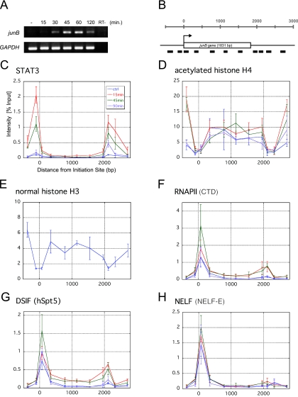
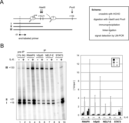
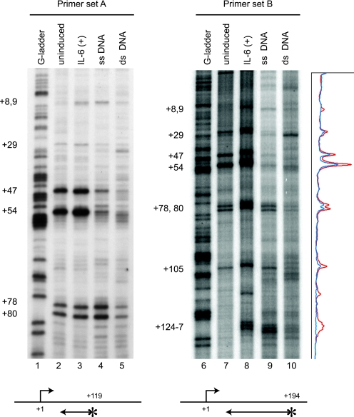
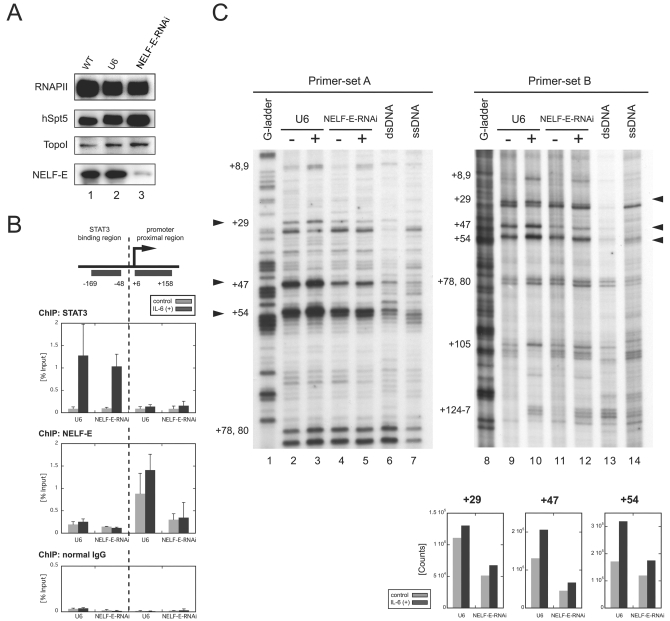
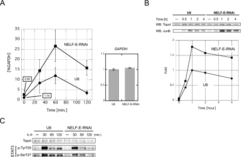
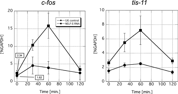
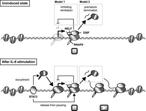
Human 5,6-dichloro-1-beta-D-ribofuranosylbenzimidazole sensitivity-inducing factor (DSIF) and negative elongation factor (NELF) negatively regulate transcription elongation by RNA polymerase II (RNAPII) in vitro. However, the physiological roles of this negative regulation are not well understood. Here, by using a number of approaches to identify protein-DNA interactions in vivo, we show that DSIF- and NELF-mediated transcriptional pausing has a dual function in regulating immediate-early expression of the human junB gene. Before induction by interleukin-6, RNAPII, DSIF, and NELF accumulate in the promoter-proximal region of junB, mainly at around position +50 from the transcription initiation site. After induction, the association of these proteins with the promoter-proximal region continues whereas RNAPII and DSIF are also found in the downstream regions. Depletion of a subunit of NELF by RNA interference enhances the junB mRNA level both before and after induction, indicating that DSIF- and NELF-mediated pausing contributes to the negative regulation of junB expression, not only by inducing RNAPII pausing before induction but also by attenuating transcription after induction. These regulatory mechanisms appear to be conserved in other immediate-early genes as well.


Figures

FIG. 1.
FIG. 1.
Spatiotemporal distribution of various factors over the junB gene. (A) RT-PCR analysis of junB gene expression. Total RNA was purified from serum-starved HepG2 cells, either left untreated or treated with 100 ng/ml IL-6 for the indicated times, and subjected to RT-PCR with two primer sets that amplify junB and GAPDH. As a control (RT−), the RT step was omitted. (B) Structure of the junB gene. Thick bars represent the positions of PCR amplicons used in ChIP assays. The transcribed region composed of a single exon is presented as an open box. (C to H) Quantification of the amount of DNA precipitated in the ChIP assay with various antibodies. HepG2 cells treated with 100 ng/ml IL-6 or left untreated were cross-linked with formaldehyde and immunoprecipitated with the indicated antibodies. Note that the histone H3 ChIP in panel E was only done on the untreated sample. Real-time PCR was carried out with the primer sets that are shown in panel B. Percent recoveries are plotted against the distance from the transcription initiation site to the midpoint of each amplicon. Data are the mean ± the standard deviation from three independent experiments, each of which was performed in duplicate.
FIG. 2.
FIG. 2.
Association of RNAPII, DSIF, and NELF with the promoter-proximal transcribed region of junB. (A) Scheme of the assay. At the left, HaeIII and PvuII cleavage sites and the position of the end-labeled primer used to detect LM-PCR products are indicated by arrows. Numbers indicate relative positions from the transcription initiation site, based on the DataBase of Transcriptional Start Sites (http://dbtss.hgc.jp/). Possible positions of RNAPII are also shown (dotted line). At the right, the experimental scheme of the assay is shown. (B) After immunoprecipitation (IP) with the indicated antibodies, coprecipitated DNA was detected by LM-PCR. Numbers at left indicate the positions of cleavage by restriction enzymes. In the graph on the right, the recoveries of fragments I, II, and III are presented as percentages of the input. Signals were quantified with a Storm 860 image analyzer (Amersham Biosciences). Data are the mean ± the standard deviation from three independent experiments.
FIG. 3.
FIG. 3.
RNAPII strongly accumulates at around position +50 of the junB gene. Control cells or cells stimulated by IL-6 for 15 min were treated with 7.5 mM KMnO4. Genomic DNA was purified, and unpaired thymine residues in transcription bubbles were mapped by in vivo footprinting with two primer sets. KMnO4 sensitivity in vivo was compared to the sensitivity of purified single-stranded (ss) or double-stranded (ds) genomic DNA. Lanes 1 and 6 contain genomic DNA partially cleaved at guanine residues. Footprinting with primer set A (lanes 1 to 5) results in higher-resolution mapping of paused sites in a smaller region than that with primer set B (lanes 6 to 10). Signal intensity of the footprinting data for primer set B was obtained with a Storm 860 image analyzer and is presented on the right. The blue and red lines represent the results of uninduced and induced states, respectively. The positions of end-labeled primers are indicated by arrows with asterisks. The numbers on the left indicate the relative positions from the transcription initiation site.
FIG. 4.
FIG. 4.
Transcriptional pausing is caused by NELF. Cells were transduced with the NELF-E-knockdown RNAi construct (NELF-E-RNAi cells) or the control U6 promoter construct (U6 cells). All experiments were performed 7 days after transduction. (A) NELF-E knockdown selectively depletes NELF-E. Whole-cell extracts were prepared from wild-type (WT) HepG2 cells, U6 cells, and NELF-E-RNAi cells and blotted with the indicated antibodies. (B) Association of NELF-E with the junB promoter-proximal region is reduced in NELF-E-RNAi cells. NELF-E-RNAi and U6 cells with or without IL-6 stimulation for 15 min were analyzed by ChIP with anti-STAT3 (top), anti-NELF-E (middle), and normal-mouse (bottom) antibodies. Recovery of the STAT3-binding region and the promoter-proximal region was quantified as for Fig. 1. Data are the mean ± the standard deviation from five (for U6) or three (NELF-E-RNAi) independent experiments. IgG, immunoglobulin G. (C) Promoter-proximal pausing is reduced in NELF-E-RNAi cells. NELF-E-RNAi and U6 cells were analyzed before and after treatment with IL-6 for 15 min by in vivo footprinting as in Fig. 3. Signal counts of the footprint data on the right at positions +29, +47, and +54 were quantified by image analyzer and are shown at the bottom. dsRNA, double-stranded RNA; ssRNA, single-stranded RNA.
FIG. 5.
FIG. 5.
NELF-E knockdown upregulates junB gene expression at the mRNA level. (A, left) junB mRNA levels in NELF-E-RNAi and U6 cells at various times after addition of IL-6. Total RNA was prepared and analyzed by real-time RT-PCR. The mRNA level of each gene was normalized against the mRNA level of GAPDH. Numerical values of uninduced states are also presented on the graph. (A, right) GAPDH mRNA levels in NELF-E-RNAi and U6 cells. Total RNA from unstimulated cells was subjected to real-time RT-PCR. The expression levels were normalized against the levels of 18S rRNA and are expressed in arbitrary units. Data obtained from three independent experiments, each performed in triplicate, are shown as the mean ± the standard deviation. (B) JunB protein levels in NELF-E-RNAi and U6 cells at various times after addition of IL-6. Whole-cell extracts were prepared and subjected to Western blot (WB) analysis with the indicated antibodies. The signal intensity of the blot was quantified with ImageJ software (http://rsb.info.nih.gov/ij/) and is expressed as n-fold change. The intensity obtained from extracts of U6 cells stimulated for 1 h was set to 1. The result was confirmed to be reproducible (data not shown). (C) Phosphorylation status of STAT3 before and after induction. Western blot analysis was performed with the indicated antibodies.
FIG. 6.
FIG. 6.
NELF-E knockdown upregulates the expression of other IEGs. The mRNA levels of c-fos and tis-11 in NELF-E-RNAi and U6 cells at various times after addition of IL-6 are shown. Total RNA was prepared and analyzed as described in the legend to Fig. 5. The mRNA level of each gene was normalized against the mRNA level of GAPDH. Data obtained from three independent experiments, in which quantifications were done in triplicate, are shown as the mean ± the standard deviation. Numerical values of uninduced states are also presented in the graph on the left.
FIG. 7.
FIG. 7.
Model for transcriptional regulation of junB by DSIF- and NELF-mediated pausing. Before induction, DSIF and NELF cause RNAPII pausing in the promoter-proximal region of junB, where chromatin structure is decondensed and permissive for PIC assembly (for details, see Discussion). After IL-6 stimulation, STAT3 binds to the promoter and 3′ enhancer regions and increases the efficiency of both PIC assembly and transcription elongation, allowing the polymerase to reach the 3′ end of the gene, possibly by recruiting P-TEFb or other proteins with elongation activation activity. However, DSIF- and NELF-mediated pausing persists in the promoter-proximal region and acts to reduce the maximal level of its expression. Two possible mechanisms for attenuation by NELF are presented.

References

    1. Abe, K., M. Hirai, K. Mizuno, N. Higashi, T. Sekimoto, T. Miki, T. Hirano, and K. Nakajima. 2001. The YXXQ motif in gp130 is crucial for STAT3 phosphorylation at Ser727 through an H7-sensitive kinase pathway. Oncogene 20:3464-3474. - PubMed
    1. Adelman, K., M. T. Marr, J. Werner, A. Saunders, Z. Ni, E. D. Andrulis, and J. T. Lis. 2005. Efficient release from promoter-proximal stall sites requires transcript cleavage factor TFIIS. Mol. Cell 17:103-112. - PubMed
    1. Aiyar, S. E., J. L. Sun, A. L. Blair, C. A. Moskaluk, Y. Z. Lu, Q. N. Ye, Y. Yamaguchi, A. Mukherjee, D. M. Ren, H. Handa, and R. Li. 2004. Attenuation of estrogen receptor alpha-mediated transcription through estrogen-stimulated recruitment of a negative elongation factor. Genes Dev. 18:2134-2146. - PMC - PubMed
    1. Andrulis, E. D., E. Guzman, P. Doring, J. Werner, and J. T. Lis. 2000. High-resolution localization of Drosophila Spt5 and Spt6 at heat shock genes in vivo: roles in promoter proximal pausing and transcription elongation. Genes Dev. 14:2635-2649. - PMC - PubMed
    1. Ausubel, F. M., R. Brent, R. E. Kingston, D. D. Moore, J. G. Seidman, J. A. Smith, and K. Struhl. 1987. Current protocols in molecular biology. John Wiley & Sons, Inc., New York, N.Y.

Publication types

MeSH terms

LinkOut - more resources